Copyright
©2014 Baishideng Publishing Group Inc.
World J Gastroenterol. Jun 14, 2014; 20(22): 6953-6960
Published online Jun 14, 2014. doi: 10.3748/wjg.v20.i22.6953
Published online Jun 14, 2014. doi: 10.3748/wjg.v20.i22.6953
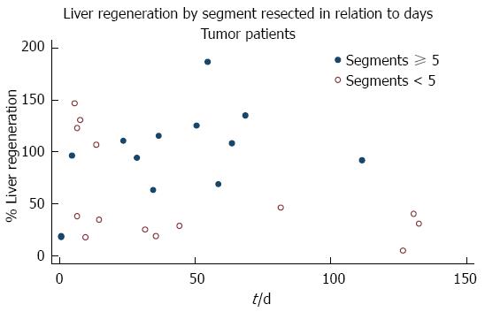
Figure 1 Distribution of liver regeneration over time in all patients.
A physiologic association between follow-up time and greater percentage of liver regeneration is showed.
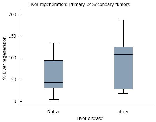
Figure 2 Box plot of liver regeneration comparing patients with primary and patients with secondary tumors.
The distribution could indicate that the type of malignancy might not influence the early liver regeneration in resected patients.
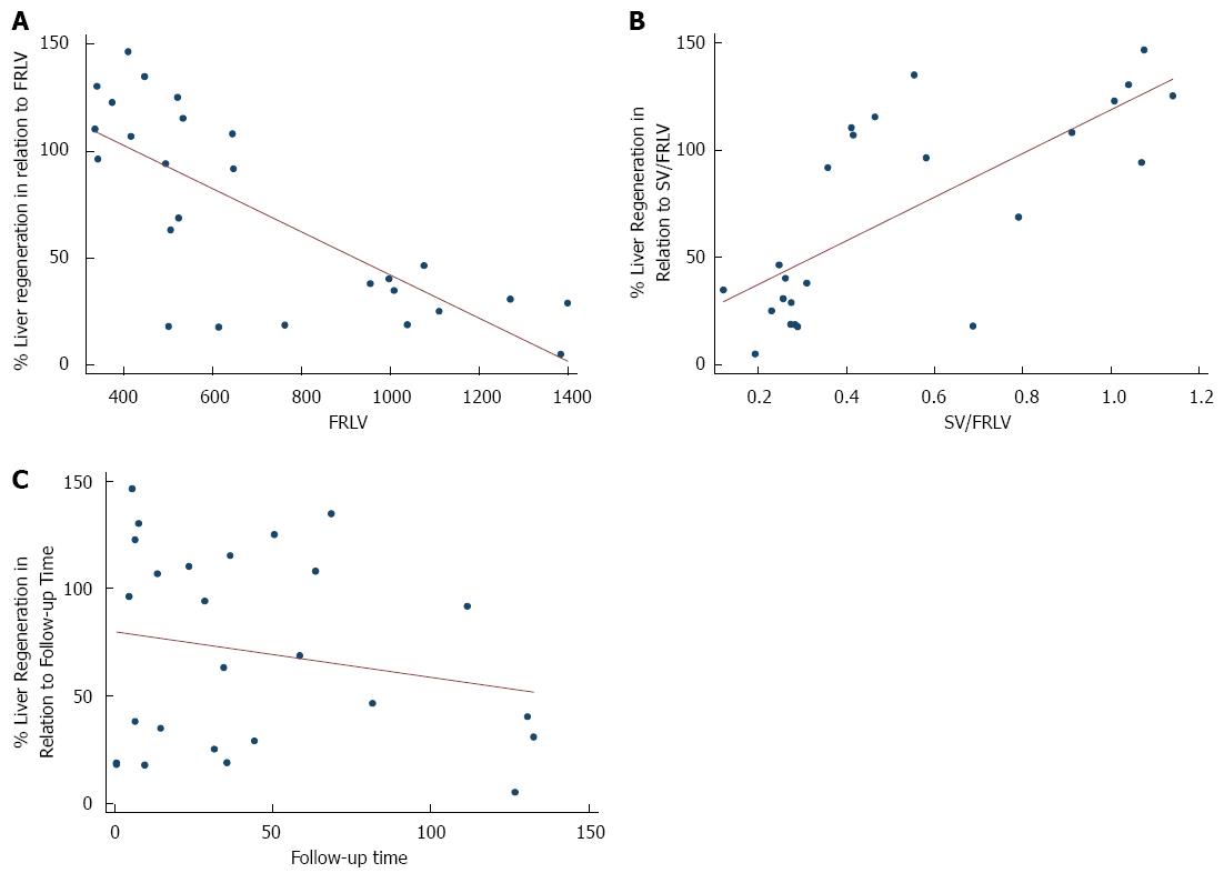
Figure 3 Scatterplot with line fit.
A: Percentage of liver regeneration in relation to future remnant liver volume (FRLV); B: Percentage of liver regeneration in relation spleen volume (SV)/FRLV; C: Percentage of liver regeneration in relation to follow-up time.
- Citation: Pagano D, Spada M, Parikh V, Tuzzolino F, Cintorino D, Maruzzelli L, Vizzini G, Luca A, Mularoni A, Grossi P, Gridelli B, Gruttadauria S. Liver regeneration after liver resection: Clinical aspects and correlation with infective complications. World J Gastroenterol 2014; 20(22): 6953-6960
- URL: https://www.wjgnet.com/1007-9327/full/v20/i22/6953.htm
- DOI: https://dx.doi.org/10.3748/wjg.v20.i22.6953


