Copyright
©2014 Baishideng Publishing Group Inc.
World J Gastroenterol. Jun 7, 2014; 20(21): 6691-6697
Published online Jun 7, 2014. doi: 10.3748/wjg.v20.i21.6691
Published online Jun 7, 2014. doi: 10.3748/wjg.v20.i21.6691
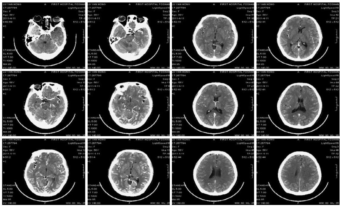
Figure 1 Brain enhanced computed tomography scan of case 1 (8 h after coma).
A mild degree of cerebral atrophy; no remarkable intracranial metastases were found.
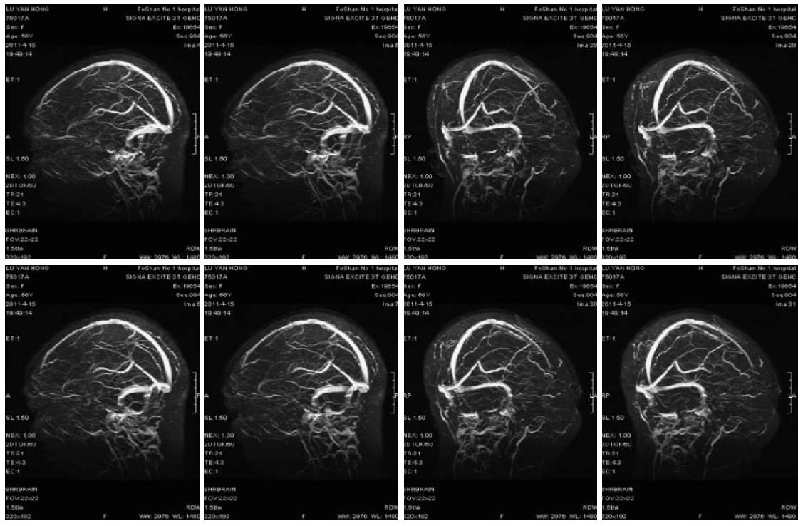
Figure 2 Enhanced brain magnetic resonance imaging and magnetic resonance venography scan of case 1 (2 d after recovery).
Mottled lesions of the left parietal lobe, indicating intracerebral microbleeds.
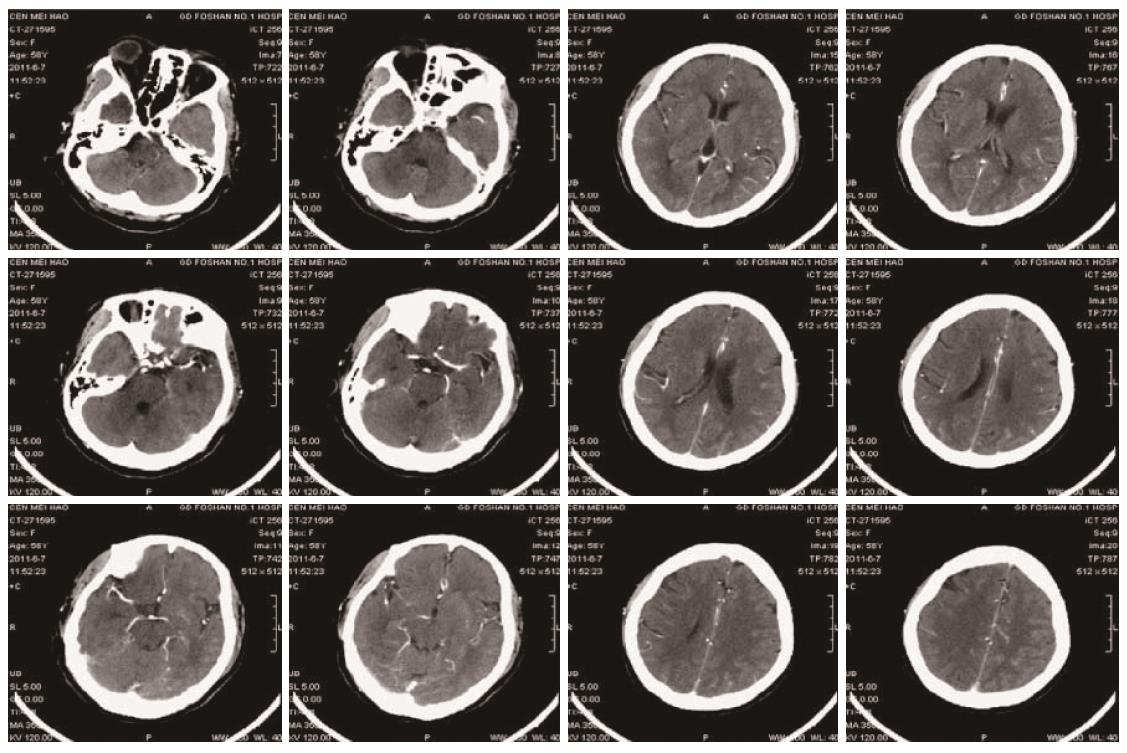
Figure 3 Enhanced computed tomography and magnetic resonance venography scan of the brain of case 2.
Mild cerebral arteriosclerosis, stenosis of the A1 segment of the right anterior cerebral artery and right intracranial artery, and effective blood supply of distal vascular and mild lacunar infarction of bilateral basal ganglia.
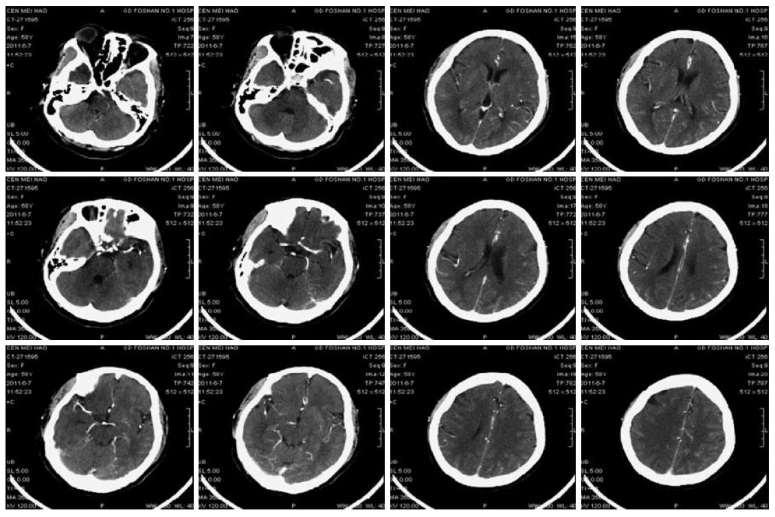
Figure 4 Repeat enhanced computed tomography and magnetic resonance venography scan of the brain of case 2 on the following day showed the same image changes as previously described.
- Citation: Wang W, Zhao LR, Lin XQ, Feng F. Reversible posterior leukoencephalopathy syndrome induced by bevacizumab plus chemotherapy in colorectal cancer. World J Gastroenterol 2014; 20(21): 6691-6697
- URL: https://www.wjgnet.com/1007-9327/full/v20/i21/6691.htm
- DOI: https://dx.doi.org/10.3748/wjg.v20.i21.6691


