Copyright
©2014 Baishideng Publishing Group Co.
World J Gastroenterol. Apr 14, 2014; 20(14): 4102-4105
Published online Apr 14, 2014. doi: 10.3748/wjg.v20.i14.4102
Published online Apr 14, 2014. doi: 10.3748/wjg.v20.i14.4102
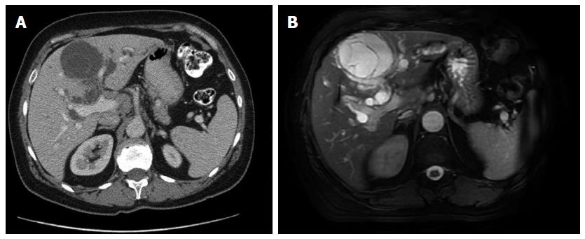
Figure 1 Computed tomography scan and T1-weighted magnetic resonance imaging.
A: Computed tomography scan (portal phase) shows a hepatic cystic tumor with thin contrast-enhanced septum and dilated intrahepatic bile ducts; B: T1-weighted magnetic resonance imaging of the tumor presents a hyperintense, multiseptal cystic lesion that does not communicate with the bile ducts.
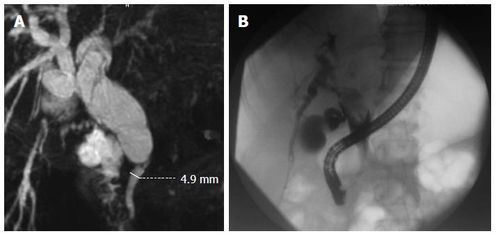
Figure 2 Magnetic resonance and preoperative endoscopic retrograde cholangiopancreatography.
A: Magnetic resonance cholangiopancreatography shows dilated main hepatic and common bile duct with a sharp change in diameter from 25 to 5 mm; B: Preoperative endoscopic retrograde cholangiopancreatography shows marked dilation of the extrahepatic biliary ducts with a contrast-filling defect caused by an oval intraductal tumor.
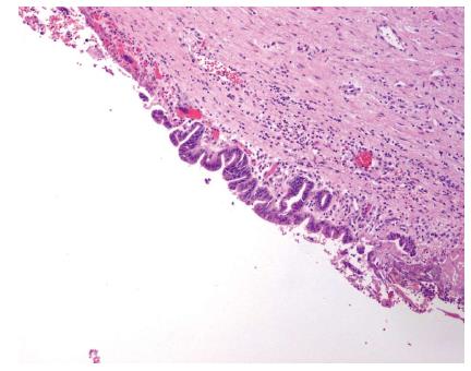
Figure 3 Histology of the intrahepatic tumor.
A: The cyst is covered with a single layer of cuboidal epithelial cells showing low grade dysplasia (hematoxylin-eosin stain, × 100 magnification); B: The tumor produces mucin; C: Ovarian-like stroma staining positively for estrogen receptor.
Figure 4 Histology of the intraductal papillary mucinous neoplasm of the bile duct showing papillary proliferation.
- Citation: Budzynska A, Hartleb M, Nowakowska-Dulawa E, Krol R, Remiszewski P, Mazurkiewicz M. Simultaneous liver mucinous cystic and intraductal papillary mucinous neoplasms of the bile duct: A case report. World J Gastroenterol 2014; 20(14): 4102-4105
- URL: https://www.wjgnet.com/1007-9327/full/v20/i14/4102.htm
- DOI: https://dx.doi.org/10.3748/wjg.v20.i14.4102


