Copyright
©The Author(s) 1996.
World J Gastroenterol. Sep 15, 1996; 2(3): 144-145
Published online Sep 15, 1996. doi: 10.3748/wjg.v2.i3.144
Published online Sep 15, 1996. doi: 10.3748/wjg.v2.i3.144
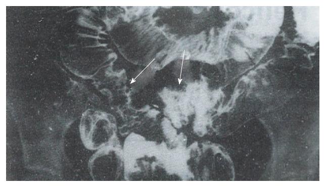
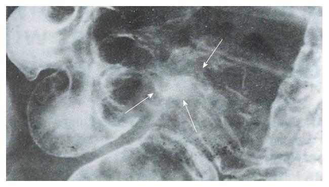
Figure 1 The mucosa is irregular and superficial ulcers (arrows) were present in several segments of the terminal ileum of TB cases.
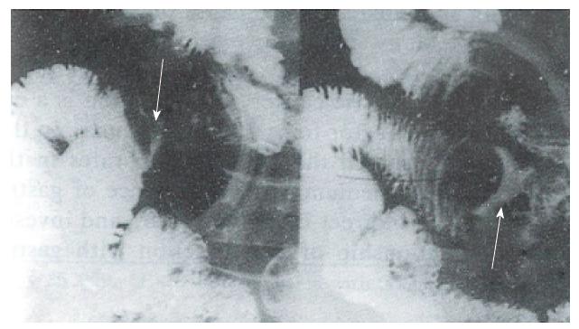
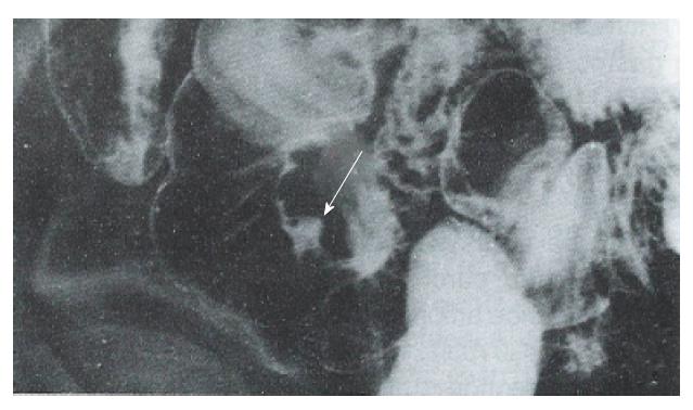
Figure 2 After compression, a transverse ulcer (arrow) is observed in a TB case.
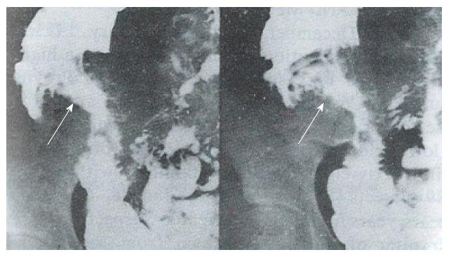
Figure 3 Triangular ulcers measuring 4 cm2 in size at the terminal ileum (arrow) were present Crohn’s disease cases.
The surrounding mucosal pattern exhibited a cobblestone appearance.
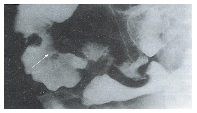
Figure 4 A round ulcer (arrow), with surrounding mild mucosal edema in the ileocecal region was present in a Crohn’s disease case.
Figure 5 A large and deep irregular ulcer (4 × 3 cm2 in size) at the terminal ileum near the ileocecal valve (double arrow) was present in a bowel Behcet disease case.
The surrounding mucosal edema (arrow) is shown.
Figure 6 A small ulcer (0.
3 × 0.4 cm2 in size) at the ileum (arrow) was present in a bowel Behcet disease case. The surrounding mucosal edema (arrow) is shown.
Figure 7 A and B: Several long, segmented, contracted, narrow, and rigid loops of the bowel in the ileum (arrow), with superficial ulcers were present in ischemic bowel disease.
No mucosal abnormalities were observed.
- Citation: Lu Y, Duan JY, Gao Y. Radiological diagnosis of inflammatory ulcerative diseases of the small bowel. World J Gastroenterol 1996; 2(3): 144-145
- URL: https://www.wjgnet.com/1007-9327/full/v2/i3/144.htm
- DOI: https://dx.doi.org/10.3748/wjg.v2.i3.144


