Copyright
©The Author(s) 1996.
World J Gastroenterol. Sep 15, 1996; 2(3): 128-130
Published online Sep 15, 1996. doi: 10.3748/wjg.v2.i3.128
Published online Sep 15, 1996. doi: 10.3748/wjg.v2.i3.128
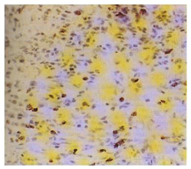
Figure 1 Poorly differentiated adenocarcinoma tissue with VEGF++ staining.
Cytoplasmic VEGF staining was seen in tumor cells and diffuse distribution of VEGF-positive tumor cells is apparent. (original magnification × 200)
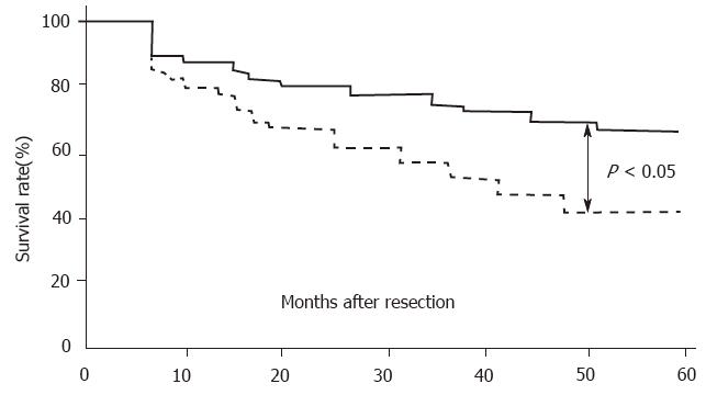
Figure 2 Survival rate after curative resection of VEGF-poor (n = 30) and VEGF-rich tumors (n = 56)
- Citation: Tao HQ, Qin LF, Lin YZ, Wang RN. Expression of vascular endothelial growth factor and its prognostic significance in gastric carcinoma. World J Gastroenterol 1996; 2(3): 128-130
- URL: https://www.wjgnet.com/1007-9327/full/v2/i3/128.htm
- DOI: https://dx.doi.org/10.3748/wjg.v2.i3.128


