Copyright
©2013 Baishideng Publishing Group Co.
World J Gastroenterol. Nov 21, 2013; 19(43): 7766-7771
Published online Nov 21, 2013. doi: 10.3748/wjg.v19.i43.7766
Published online Nov 21, 2013. doi: 10.3748/wjg.v19.i43.7766
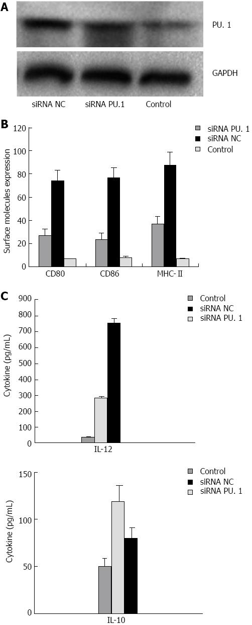
Figure 1 Identification of PU.
1-silenced dendritic cells in vitro. A: Analyses of PU.1 gene expression by Western blot; B: Flow cytometry analysis of CD80, CD86 and major histocompatibility complex (MHC)-II in dendritic cells; C: Analyses of cell culture supernatants by enzyme-linked immunosorbent assay. CD: Cluster of differentiation; GAPDH: Reduced glyceraldehyde-phosphate dehydrogenase.
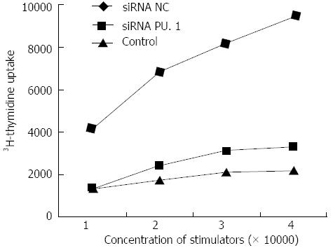
Figure 2 PU.
1-silenced dendritic cells suppress allogeneic T cell proliferation. Bone marrow dendritic cells (DCs) were cultured and transfected with PU.1 siRNA as described. After that, three groups of DCs were collected and co-cultured with allogeneic T cells in a 96-well plate at various ratios as indicated. [3H] was added 48 h after coculture, and its incorporation was measured as an indicator of T cell proliferation (P < 0.05 siRNA PU.1 vs siRNA NC group).
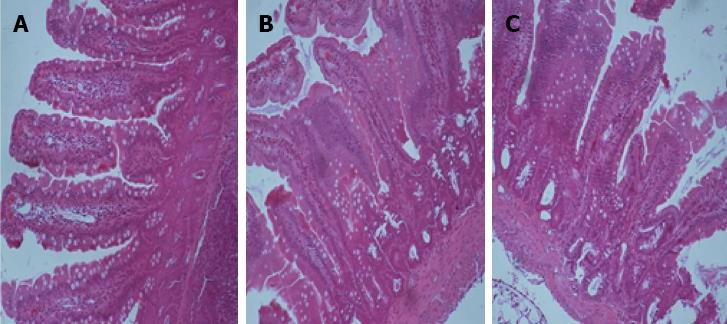
Figure 3 Histopathology of intestinal allograft from recipient rats.
Treatment with PU.1 small interference RNA (siRNA) prevents allograft rejection and is associated with mild lymphocyte infiltration and villous damage. Samples from the three groups were compared (magnification × 40, scale bar 20 μm). A: The siRNA PU.1 group; B: The siRNA NC group; C: The control group.
- Citation: Xu XW, Ding BW, Zhu CR, Ji W, Li JS. PU.1-silenced dendritic cells prolong allograft survival in rats receiving intestinal transplantation. World J Gastroenterol 2013; 19(43): 7766-7771
- URL: https://www.wjgnet.com/1007-9327/full/v19/i43/7766.htm
- DOI: https://dx.doi.org/10.3748/wjg.v19.i43.7766


