Copyright
©2013 Baishideng Publishing Group Co.
World J Gastroenterol. Nov 21, 2013; 19(43): 7758-7765
Published online Nov 21, 2013. doi: 10.3748/wjg.v19.i43.7758
Published online Nov 21, 2013. doi: 10.3748/wjg.v19.i43.7758
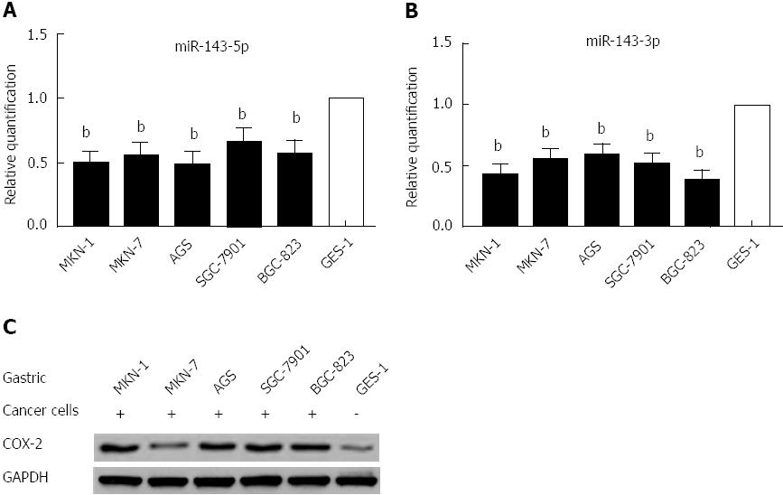
Figure 1 MicroRNA-143 expression is downregulated in gastric cancer cell lines.
A: Quantitative real-time polymerase chain reaction analysis was performed in five gastric cell lines and a normal gastric epithelium cell line (GES-1). Mature microRNA-143-5p (miR-143-5p) expression levels were significantly downregulated in gastric cancer cells, compared to normal gastric epithelium cells (bP < 0.01). The mean value from the GES-1 cell line was normalized to 1; B: Mature miR-143-3p expression levels were significantly downregulated in gastric cancer cells, compared to normal gastric epithelium cells (bP < 0.01); C: Western blot analysis showed that cyclooxygenase-2 protein expression in the five human gastric cancer cell lines inversely correlated with the miR-143 levels. GAPDH: Glyceraldehyde-3-phosphate dehydrogenase; COX-2: Cyclooxygenase-2.
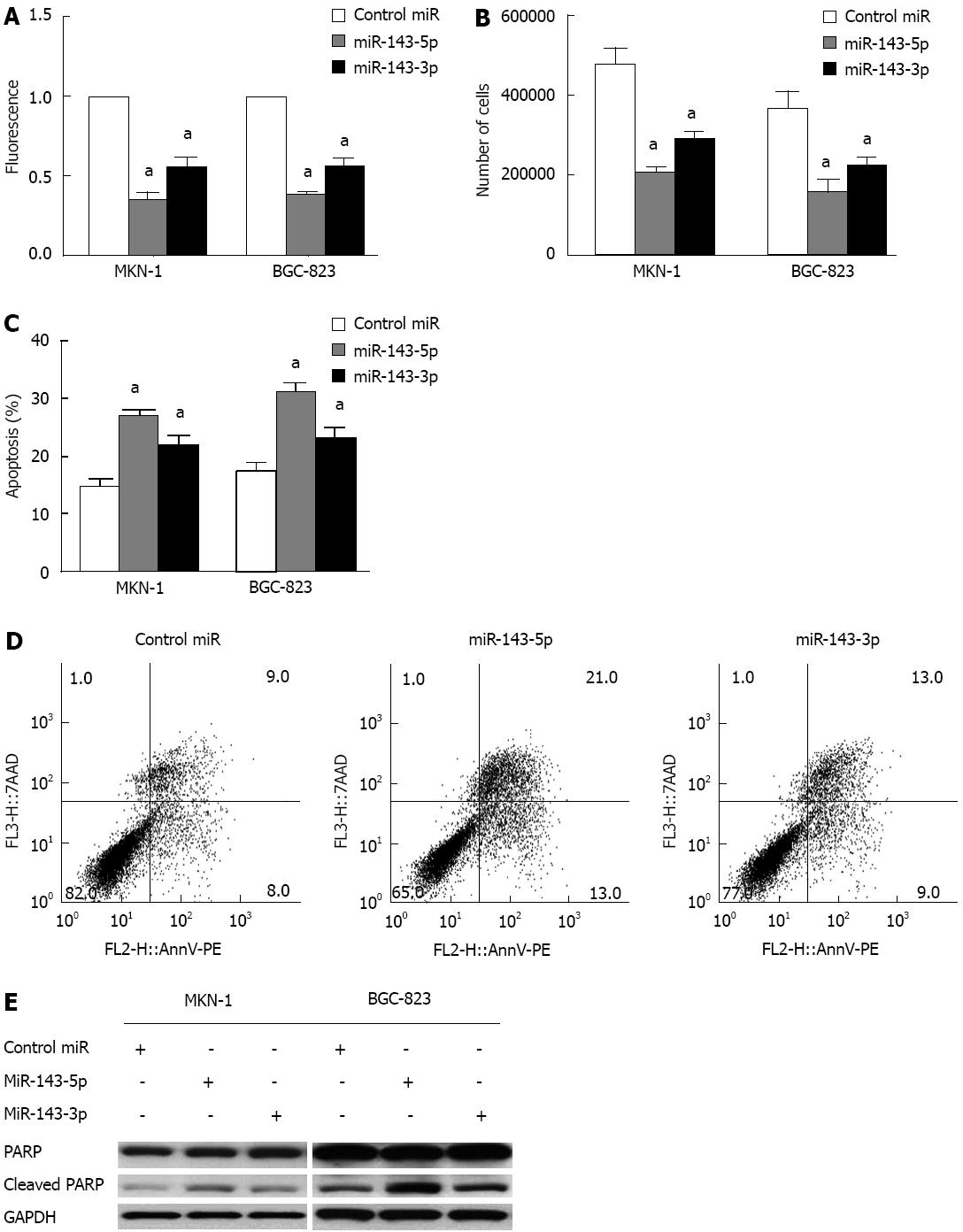
Figure 2 Transfection with microRNA-143 inhibits gastric cancer cell viability and induces apoptosis.
A: An Alamar Blue assay was performed 3 d after transfection with microRNA-143-5p (miR-143-5p) or miR-143-3p to measure the viability of MKN-1 and BGC-823 gastric cancer cells. The results showed significant decreases in cell viability following transfection with either miR-143-5p or miR-143-3p (aP < 0.05); this decrease was greater after miR-143-5p transfection than after miR-143-3p (cP < 0.05); B: Cell counts performed 3 d after transfection into MKN-1 and BGC-823 gastric cancer cells showed decreased cell numbers after transfection with either miR-143-5p or miR-143-3p (aP < 0.05). Consistent with the results of the Alamar Blue assay, the decrease in cell number after miR-143-5p transfection was greater than that after miR-143-3p transfection (cP < 0.05); C and D: An Annexin V/PE cell apoptosis assay revealed increased apoptosis in gastric cancer cells after transfection with either miR-143-5p or miR-143-3p (aP < 0.05). Cells transfected with miR-143-5p had a significantly higher apoptosis rate than those transfected with miR-143-3p (cP < 0.05); E: Western blots of PARP protein in MKN-1 and BGC-823 cell lines at 3 d after transfection with miR-143-5p or miR-143-3p. GAPDH was used as a control. The results showed increased expression of cleaved PARP after miR-143-5p and miR-143-3p transfection. GAPDH: Glyceraldehyde-3-phosphate dehydrogenase. PARP: Poly (ADP-ribose) polymerase.
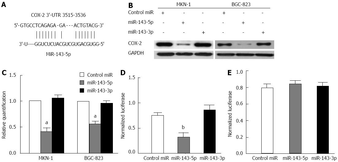
Figure 3 MicroRNA-143-5p directly inhibits cyclooxygenase-2 expression.
A: Sites of miR-143-5p seed matches in the cyclooxygenase-2 (COX-2) 3′-untranslated region (3′-UTR) (nucleotides 3515-3536); B: Western blot of COX-2 protein in the MKN-1 and BGC-823 gastric cancer cell lines at 3 d after transfection with miR-143-5p, miR-143-3p or microRNA (miRNA) mimic control. GAPDH was used as a control. The results showed a profound decrease in COX-2 protein expression after transfection with miR-143-5p but not miR-143-3p; C: Real-time reverse transcription-polymerase chain reaction to determine COX-2 mRNA expression was performed 2 d after transfection with miR-143-5p, miR-143-3p or a control in MKN-1 and BGC-823. The mean expression in the control group was normalized to 1. Consistent with transcriptional inhibition by miRNA, the COX-2 mRNA level was reduced after miR-143-5p transfection (aP < 0.05); D: Normalized activity of the wild-type COX-2 3′-UTR luciferase reporter in BGC-823 cells, 2 d after transfection with miR-143-5p, miR-143-3p or a control. The luciferase activity was significantly decreased by miR-143-5p (bP < 0.01) but not miR-143-3p; E: Normalized activity of the mutant-type COX-2 3′-UTR luciferase reporter in BGC-823 cells, 2 d after transfection with miR-143-5p, miR-143-3p or a control. The results showed that cotransfection of the mutant reporter plasmid with miR-143-5p or miR-143-3p had no effect on luciferase activity in the transfected cells. GAPDH: Glyceraldehyde-3-phosphate dehydrogenase.
- Citation: Wu XL, Cheng B, Li PY, Huang HJ, Zhao Q, Dan ZL, Tian DA, Zhang P. MicroRNA-143 suppresses gastric cancer cell growth and induces apoptosis by targeting COX-2. World J Gastroenterol 2013; 19(43): 7758-7765
- URL: https://www.wjgnet.com/1007-9327/full/v19/i43/7758.htm
- DOI: https://dx.doi.org/10.3748/wjg.v19.i43.7758


