Copyright
©2013 Baishideng Publishing Group Co.
World J Gastroenterol. Jan 28, 2013; 19(4): 492-502
Published online Jan 28, 2013. doi: 10.3748/wjg.v19.i4.492
Published online Jan 28, 2013. doi: 10.3748/wjg.v19.i4.492
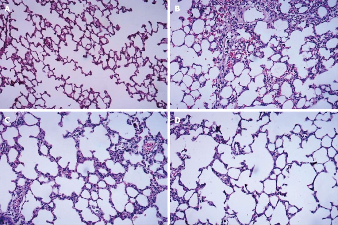
Figure 1 Hematoxylin and eosin staining of dorsal lobe of right lung tissue.
A: Control group; B: Septic shock group; C: The early fluid resuscitation-treated group; D: Combined early fluid resuscitation and 2% hydrogen inhalation-treated group. Lung tissues were fixed with 4% paraformaldehyde for more than 24 h. After dehydrating and embedding, they were cut into 5 μm slices, and were stained with hematoxylin and eosin. The pathogenesis of lung injury was observed under a microscope (original magnification × 200). Normal alveolar structure was found in Group A. In Group B, disorders of the alveolar structures, severe neutrophil infiltration in the alveoli and alveolar capillary congestion were observed. In Group C, the extent of neutrophil accumulation and the alveolar-capillary exudate were reduced compared with Group B. However, significant decreases in alveolar damage were found in Group D, and there was significant improvement in alveolar edema compared with Group C.
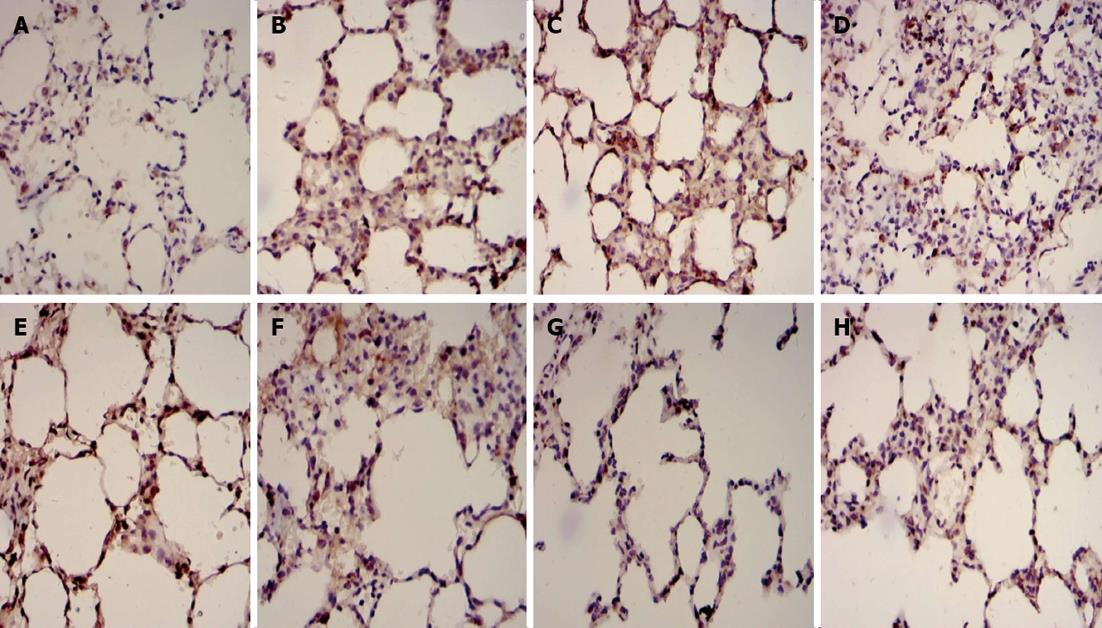
Figure 2 Immunohistochemical examples for Fas and Bcl2 in dorsal lobe of right lung tissue.
A: Fas in control group; B: Fas in septic shock group; C: Fas in early fluid resuscitation-treated group; D: Fas in early fluid resuscitation + 2% hydrogen inhalation-treated group; E: Bcl2 in control group; F: Bcl2 in septic shock group; G: Bcl2 in early fluid resuscitation-treated group; H: Bcl2 in early fluid resuscitation + 2% hydrogen inhalation-treated group. Examples were examined under a light microscope equipped with an image analysis system. Yellow or brownish-yellow staining represents positive staining. The expression of Fas proteins was up-regulated, while the expression of Bcl2 proteins was down-regulated in Group B compared with Group A. In Group C, however, the expression of Fas proteins was down-regulated, and the expression of Bcl2 proteins was up-regulated compared with Group B. In addition, the lower expression of Fas proteins and the higher expression of Bcl2 proteins were observed in Group D compared with Group C.
Figure 3 Levels of Fas and Bcl2 proteins in dorsal lobe of right lung tissues quantified by Western blotting.
β-actin was used as a loading control. The relative Fas (A) and Bcl2 (B) protein levels of lung tissues from the four groups were expressed as arbitrary units after normalization with β-actin (C: Fas; D: Bcl2). Data were considered significant at aP < 0.05 and highly significant at bP < 0.01 vs the control group. The level of Fas protein was up-regulated, while the level of Bcl2 protein was down-regulated in Group B vs Group A. However, in Group C the level of Fas proteins was down-regulated, and the level of Bcl2 proteins was up-regulated vs Group B (P < 0.01). In addition, the lower level of Fas protein and the higher level of Bcl2 protein was found in Group D compared with Group C (P < 0.05).
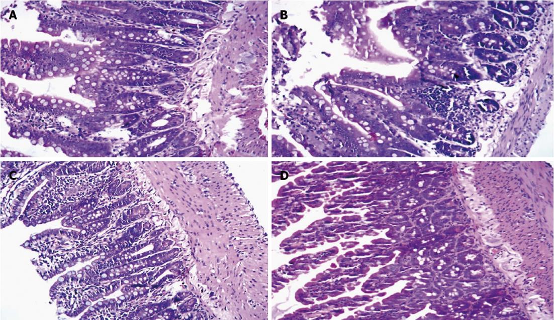
Figure 4 Hematoxylin and eosin staining of small intestine tissue.
A: Control group; B: Septic shock group; C: Early fluid resuscitation-treated group; D: Early fluid resuscitation + 2% hydrogen inhalation-treated group. Small intestinal tissues were fixed with 4% paraformaldehyde for more than 24 h. After dehydrating and embedding, they were cut into 5 μm slices, and were stained with hematoxylin and eosin. The pathogenesis of small intestine injury was observed under a light microscope (original magnification × 200). In Group A, normal intestinal mucosa was observed. In Group B, glands of small intestine were significantly damaged, and severe edema of mucosal villi, neutrophil infiltration, epithelial cell sloughing off, and even small bowel ulceration were observed. The damage mentioned above was far more severe in Group C. But in Group D, the damage was significantly reduced.
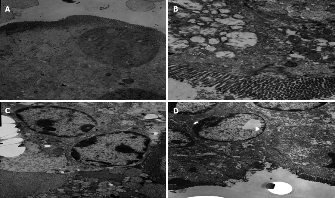
Figure 5 Changes in the organelles of small intestinal tissues under transmission electron microscope.
A: Control group; B: Septic shock group; C: Early fluid resuscitation-treated group; D: Early fluid resuscitation + 2% hydrogen inhalation-treated group. The changes in the organelles were observed under transmission electron microscope. In Group A, normal microvilli on the surface of epithelial cells of the intestine were observed. In Group B, intestinal microvillus reduction was observed. Mitochondria showed vacuolization and heterochromatin nuclei showed margination phenomena. The microvilli on the surface of epithelial cells of the intestine were sparse in Group C, and reduction of the cristae of mitochondria, an abundance of marginated heterochromatin nuclei, and severe rough endoplasmic reticulum swelling and expansion were observed. In Group D, the microvilli were missing to a small extent, and the heterochromatin nuclei showed only mild margination.
- Citation: Liu W, Shan LP, Dong XS, Liu XW, Ma T, Liu Z. Combined early fluid resuscitation and hydrogen inhalation attenuates lung and intestine injury. World J Gastroenterol 2013; 19(4): 492-502
- URL: https://www.wjgnet.com/1007-9327/full/v19/i4/492.htm
- DOI: https://dx.doi.org/10.3748/wjg.v19.i4.492


