Copyright
©2013 Baishideng Publishing Group Co.
World J Gastroenterol. Jan 28, 2013; 19(4): 492-502
Published online Jan 28, 2013. doi: 10.3748/wjg.v19.i4.492
Published online Jan 28, 2013. doi: 10.3748/wjg.v19.i4.492
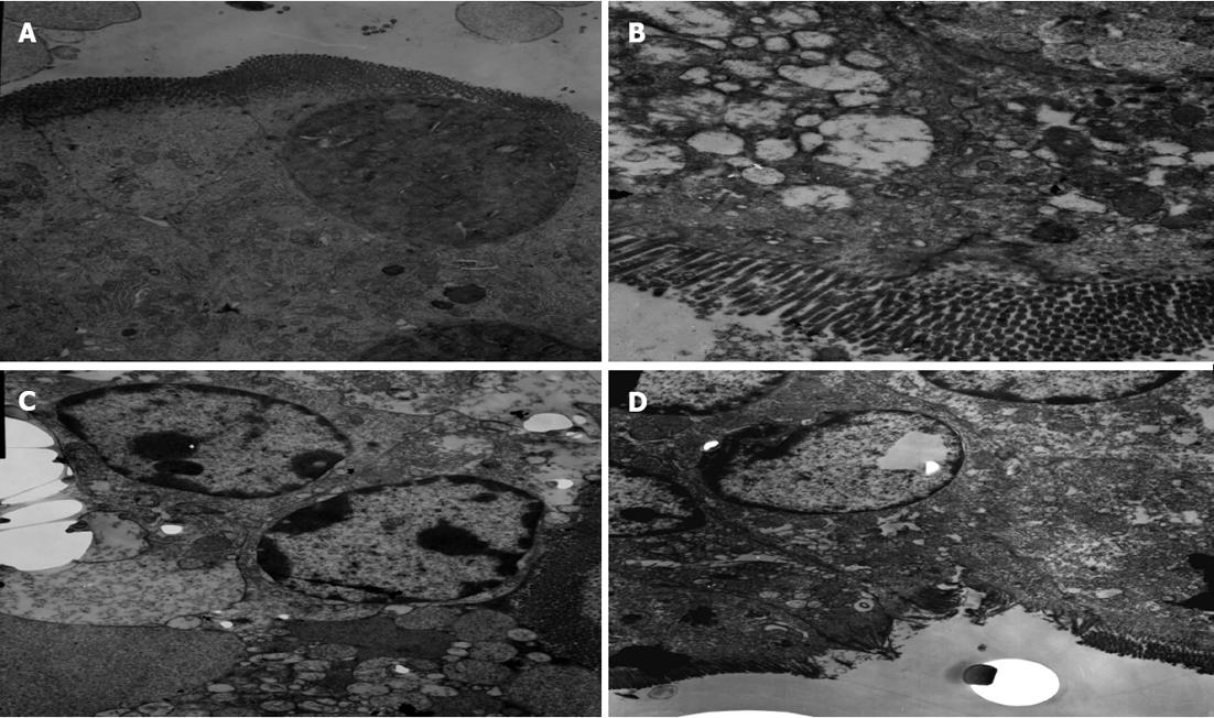
Figure 5 Changes in the organelles of small intestinal tissues under transmission electron microscope.
A: Control group; B: Septic shock group; C: Early fluid resuscitation-treated group; D: Early fluid resuscitation + 2% hydrogen inhalation-treated group. The changes in the organelles were observed under transmission electron microscope. In Group A, normal microvilli on the surface of epithelial cells of the intestine were observed. In Group B, intestinal microvillus reduction was observed. Mitochondria showed vacuolization and heterochromatin nuclei showed margination phenomena. The microvilli on the surface of epithelial cells of the intestine were sparse in Group C, and reduction of the cristae of mitochondria, an abundance of marginated heterochromatin nuclei, and severe rough endoplasmic reticulum swelling and expansion were observed. In Group D, the microvilli were missing to a small extent, and the heterochromatin nuclei showed only mild margination.
- Citation: Liu W, Shan LP, Dong XS, Liu XW, Ma T, Liu Z. Combined early fluid resuscitation and hydrogen inhalation attenuates lung and intestine injury. World J Gastroenterol 2013; 19(4): 492-502
- URL: https://www.wjgnet.com/1007-9327/full/v19/i4/492.htm
- DOI: https://dx.doi.org/10.3748/wjg.v19.i4.492


