Copyright
©2013 Baishideng Publishing Group Co.
World J Gastroenterol. Aug 21, 2013; 19(31): 5178-5181
Published online Aug 21, 2013. doi: 10.3748/wjg.v19.i31.5178
Published online Aug 21, 2013. doi: 10.3748/wjg.v19.i31.5178
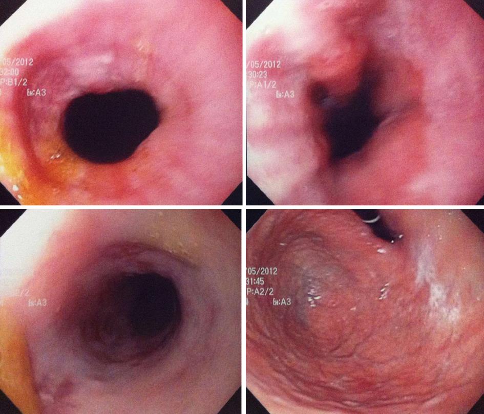
Figure 1 Stenosis of the esophagus, day 4.
Figure 2 Hyperhemia consistent with esophagitis with complete resolution of the stenosis of the esophagus, day 8.
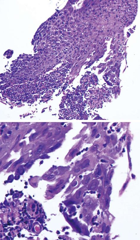
Figure 3 Ulcerated squamous epithelium with herpes virus inclusions easily visible on hematoxylin phloxine saffron coloration.
- Citation: Jetté-Côté I, Ouellette D, Béliveau C, Mitchell A. Total dysphagia after short course of systemic corticotherapy: Herpes simplex virus esophagitis. World J Gastroenterol 2013; 19(31): 5178-5181
- URL: https://www.wjgnet.com/1007-9327/full/v19/i31/5178.htm
- DOI: https://dx.doi.org/10.3748/wjg.v19.i31.5178


