Copyright
©2013 Baishideng Publishing Group Co.
World J Gastroenterol. Jul 14, 2013; 19(26): 4257-4261
Published online Jul 14, 2013. doi: 10.3748/wjg.v19.i26.4257
Published online Jul 14, 2013. doi: 10.3748/wjg.v19.i26.4257
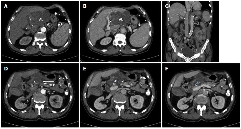
Figure 1 Radiological findings prior and after curative resection.
A, B: Computed tomography of the abdomen displaying an extended tumour manifestation prior to resection; C: Computed tomography of the abdomen following extended left hemihepatectomy; D-F: Computed tomography of the abdomen prior to resection. Arrows: possible extensive adhesions to adjacent pancreatic head and corpus. AE: Alveolar echinococcus tumour; P: Pancreas.
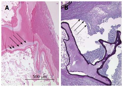
Figure 2 Referral evaluations for diagnosis of alveolar echinococcosis.
A: The hematoxylin and eosin stain of paraffin sections displays the laminated layer as a narrow band (long arrows). The germinal layer is marked by short arrows; B: Periodic acid-Schiff (PAS) stain shows a strongly PAS-positive basophilic laminated layer displaying a bizarre narrow structural pattern. The long arrows indicate the typical severe inflammatory process associated with the characteristic tubular growth pattern of the parasite.
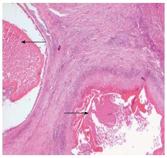
Figure 3 Histological findings after curative resection.
Hematoxylin and eosin stain of paraffin sections displaying two daughter cysts containing no vital protoscoleces embedded in a larger lesion. Black arrows indicate avital protoscoleces.
- Citation: Atanasov G, Benckert C, Thelen A, Tappe D, Frosch M, Teichmann D, Barth TF, Wittekind C, Schubert S, Jonas S. Alveolar echinococcosis-spreading disease challenging clinicians: A case report and literature review. World J Gastroenterol 2013; 19(26): 4257-4261
- URL: https://www.wjgnet.com/1007-9327/full/v19/i26/4257.htm
- DOI: https://dx.doi.org/10.3748/wjg.v19.i26.4257


