Copyright
©2013 Baishideng Publishing Group Co.
World J Gastroenterol. Jul 14, 2013; 19(26): 4257-4261
Published online Jul 14, 2013. doi: 10.3748/wjg.v19.i26.4257
Published online Jul 14, 2013. doi: 10.3748/wjg.v19.i26.4257
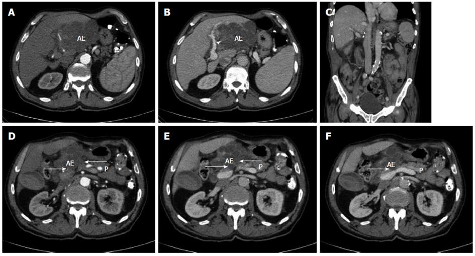
Figure 1 Radiological findings prior and after curative resection.
A, B: Computed tomography of the abdomen displaying an extended tumour manifestation prior to resection; C: Computed tomography of the abdomen following extended left hemihepatectomy; D-F: Computed tomography of the abdomen prior to resection. Arrows: possible extensive adhesions to adjacent pancreatic head and corpus. AE: Alveolar echinococcus tumour; P: Pancreas.
- Citation: Atanasov G, Benckert C, Thelen A, Tappe D, Frosch M, Teichmann D, Barth TF, Wittekind C, Schubert S, Jonas S. Alveolar echinococcosis-spreading disease challenging clinicians: A case report and literature review. World J Gastroenterol 2013; 19(26): 4257-4261
- URL: https://www.wjgnet.com/1007-9327/full/v19/i26/4257.htm
- DOI: https://dx.doi.org/10.3748/wjg.v19.i26.4257


