Copyright
©2013 Baishideng Publishing Group Co.
World J Gastroenterol. Apr 21, 2013; 19(15): 2331-2339
Published online Apr 21, 2013. doi: 10.3748/wjg.v19.i15.2331
Published online Apr 21, 2013. doi: 10.3748/wjg.v19.i15.2331
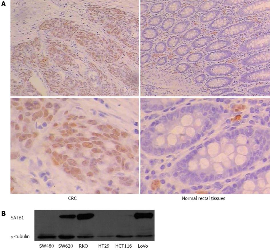
Figure 1 Upregulation of special AT-rich sequence-binding protein 1 in human colorectal cancer.
A: Immunostaining for SATB1 protein in tissue of carcinomas and adjacent normal tissue mucosa. Top: Representative pictures of carcinomas (left) and normal tissue (right); B: Western blot detection of SATB1 protein in different colorectal cancer cell lines. SATB1: Special AT-rich sequence-binding protein 1; CRC: Colorectal cancer.
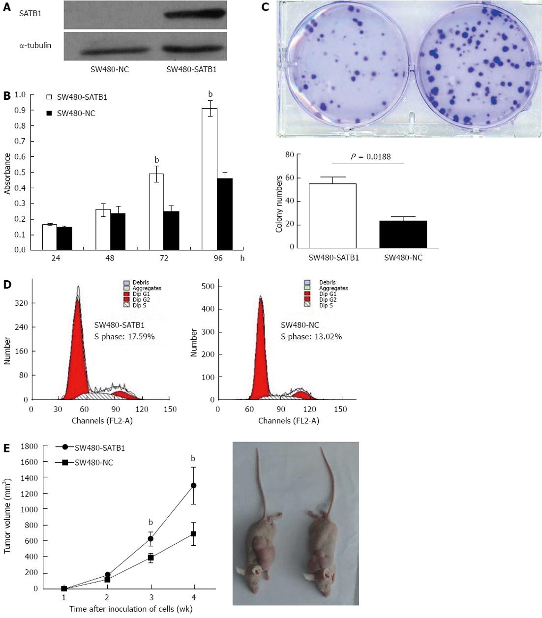
Figure 2 Special AT-rich sequence-binding protein 1 promotes cell growth and in vivo tumorigenesis potential in SW480 cells.
A: Western blot analysis of special AT-rich sequence-binding protein 1 (SATB1) protein expression in colorectal cancer (CRC) cells that stably expressing SATB1 and SW480 negative control (NC) cells; B: Effect of SATB1 overexpression on cell proliferation by 3-(4,5-cimethylthiazol-2-yl)-2,5-diphenyl tetrazolium bromide assay. bP < 0.01 vs NC cells; C: Colony formation assays for SW480-SATB1 cells and NC cells. Data are representatives of three independent experiments; D: Growth rates of SW480-SATB1 cells and negative control cells in an in vivo mouse model. Volumes of tumors were monitored every week. Bottom: Representative pictures of tumor samples. bP < 0.01 vs NC cells.
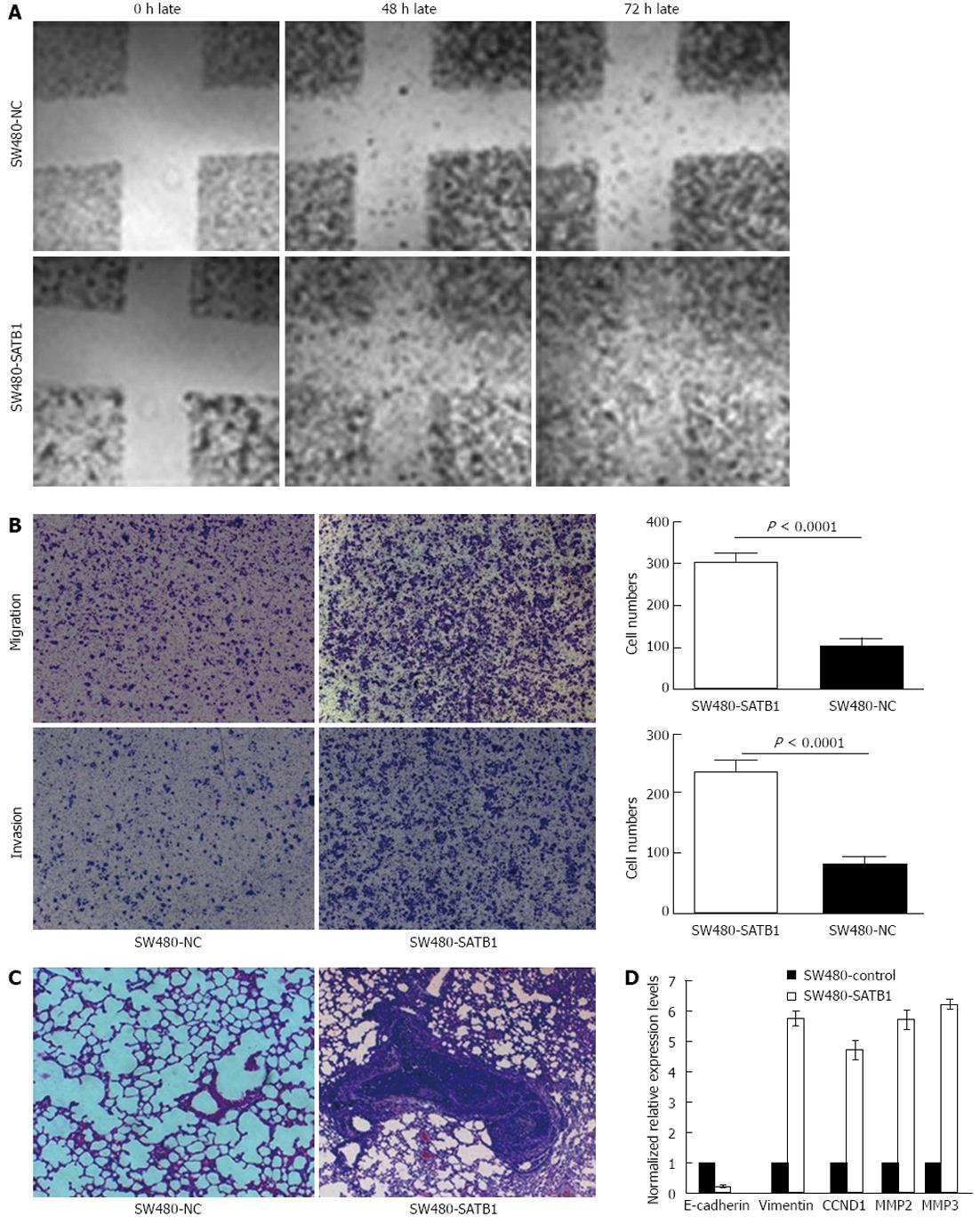
Figure 3 Special AT-rich sequence-binding protein 1 promotes migratory property of SW480 cell in vitro and in vivo metastasis potential.
A: A wound-healing assay of SW480-special AT-rich sequence-binding protein 1 (SATB1) cells and negative control (NC) cells. Photographs were taken at the time of 0, 48 and 72 h. Representative photos from one of three replicate experiments are shown (40× original magnification); B: Representative photo-micrographs of Transwell results for SW480-SATB1 cells and NC cells were taken (40× original magnification). The number of SW480-SATB1 cells passing through the membrane with or without Matrigel was significantly lower than that of NCs; C: Representative hematoxylin and eosin stained sections of the lung tissues isolated from mice implanted with SW480-SATB1 cells and NC cells through the lateral tail vein. The data shown are the number of lung metastases from each group; D: A bar chart showing downstream molecules regulated by SATB1 using real-time polymerase chain reaction in SW480-SATB1 cells and NC cells. MMP: Matrix metalloproteases; CCND1: Cyclin D1.
- Citation: Fang XF, Hou ZB, Dai XZ, Chen C, Ge J, Shen H, Li XF, Yu LK, Yuan Y. Special AT-rich sequence-binding protein 1 promotes cell growth and metastasis in colorectal cancer. World J Gastroenterol 2013; 19(15): 2331-2339
- URL: https://www.wjgnet.com/1007-9327/full/v19/i15/2331.htm
- DOI: https://dx.doi.org/10.3748/wjg.v19.i15.2331


