Copyright
©2013 Baishideng Publishing Group Co.
World J Gastroenterol. Apr 14, 2013; 19(14): 2197-2207
Published online Apr 14, 2013. doi: 10.3748/wjg.v19.i14.2197
Published online Apr 14, 2013. doi: 10.3748/wjg.v19.i14.2197
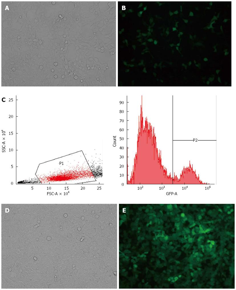
Figure 1 SW-620 cells transduced by lentivirus before and after flow cytometry selection.
A, B: SW-620 cells transduced by lentivirus before flow cytometry selection (A: Light microscopy; B: Fluorescent microscopy × 40); C: SW-620 cells with green fluorescent protein+ were distinguished by flow cytometry; D, E: SW-620 cells transduced by lentivirus after flow cytometry selection (D: Light microscopy; E: Fluorescent microscopy × 40).
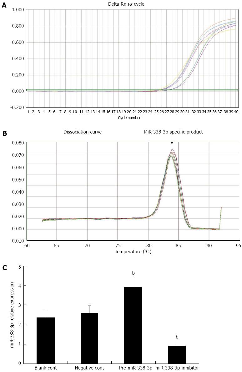
Figure 2 Real-time reverse transcriptase-polymerase chain reaction analysis detecting miRNA-338-3p expression in SW-620 cells.
A: miRNA-338-3p (miR-338-3p) cDNA concentrations, Log value as ordinate, Ct value as abscissa; B: Tm of miR-338-3p was 84.09 °C; C: Expression of miR-338-3p detected by real-time reverse transcriptase-polymerase chain reaction. Expression of U6 snRNA was used as an internal control. bP < 0.01 vs control group.
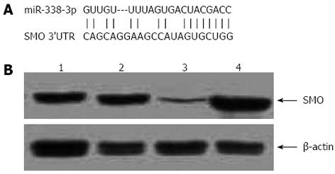
Figure 3 miRNA-338-3p regulates expression of smoothened in SW-620 cells.
A: Smoothened (SMO) 3’-untranslated region potentially targeted by miRNA-338-3p (miR-338-3p) as predicted by TargetScan; B: Western blotting analysis showing SMO protein expression in SW-620 cells. β-actin was used as a housekeeping gene to normalize SMO protein expression. Lane 1: blank control; lane 2: SW-620 cells transduced with lentivirus pLV-THM-control; lane 3: SW-620 cells transduced with lentivirus pLV-THM-miR-338-3p; lane 4: SW-620 cells transduced with lentivirus pLV-THM-miR-338-3p-inhibitor. The results are representative of three independent experiments.
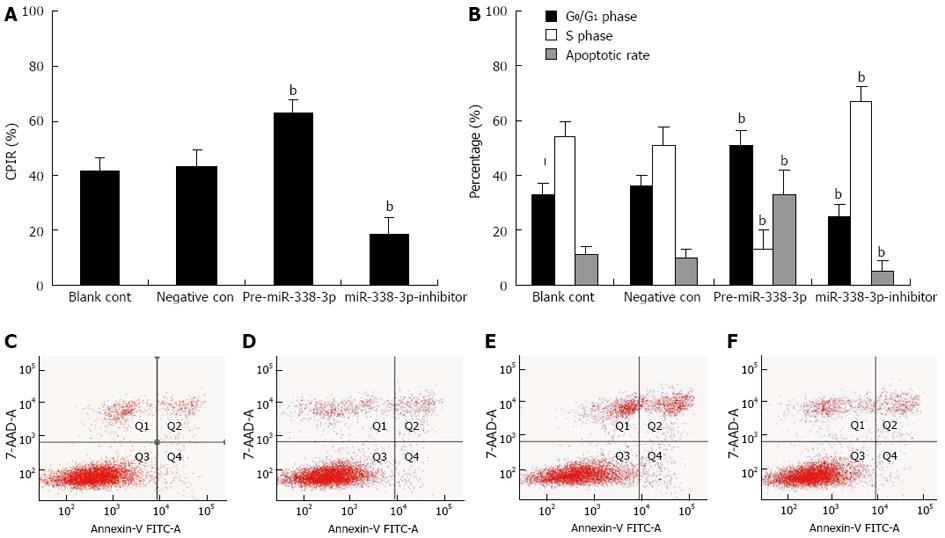
Figure 4 Effects of miRNA-338-3p on cell proliferation and apoptosis in colorectal carcinoma cells.
A: Cell proliferation was determined by 3-(4,5-dimethyl-2 thiazoyl)-2,5-diphenyl-2H-tetrazolium bromide assay. Cellular proliferation inhibition rate (CPIR) in the presence of pre-miRNA-338-3p (miR-338-3p) or miR-338-3p-inhibitor was compared with that of the controls; n = 6, mean ± SD. bP < 0.01 vs control group; B: Effects of pre-miR-338-3p and miR-338-3p-inhibitor on cell-cycle in SW-620 cells. The percentages of cells in G0/G1 phase and S phase and apoptotic rate were measured by computing the ratio of the number of corresponding cells to total cells; n = 3, mean ± SD. bP < 0.01 vs control group; C-F: Apoptosis analysis of transduced cells by flow cytometry. C: Blank control; D: SW-620 cells transduced with lentivirus pLV-THM-control; E: SW-620 cells transduced with lentivirus pLV-THM-miR-338-3p; F: SW-620 cells transduced with lentivirus pLV-THM-miR-338-3p-inhibitor. The right lower quadrant (FITC+/PI-) shown as apoptotic cells.
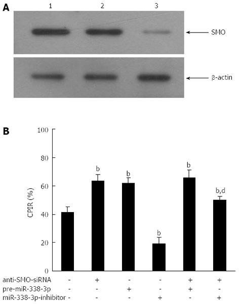
Figure 5 Ectopic expression of miRNA-338-3p affects proliferation of colorectal carcinoma cells by targeting smoothened.
SW-620 cells were pretreated with or without anti-smoothened (SMO)-siRNA (50 nmol/L) for 24 h prior to transduction with lentivirus pLV-THM-miRNA-338-3p (miR-338-3p) or pLV-THM-miR-338-3p-inhibitor. A: Western blotting analysis showing that SMO protein reduced markedly after transfection with anti-SMO-siRNA. Equal loading was confirmed by using β-actin. Lane 1, blank control; lane 2, SW-620 cells transfected with control siRNA; lane 3, SW-620 cells transfected with anti-SMO-siRNA; B: Cell proliferation was determined by 3-(4,5-dimethyl-2 thiazoyl)-2,5-diphenyl-2H-tetrazolium bromide assay. Enhancement of SW-620 cell proliferation by miR-338-3p-inhibitor was largely, but not completely, abrogated by anti-SMO-siRNA [cellular proliferation inhibition rate (CPIR) from 19.2% to 50.9%]; n = 3, mean ± SD. bP < 0.01 vs negative control group. dP < 0.01 vs sole miR-338-3p-inhibitor group.
- Citation: Sun K, Deng HJ, Lei ST, Dong JQ, Li GX. miRNA-338-3p suppresses cell growth of human colorectal carcinoma by targeting smoothened. World J Gastroenterol 2013; 19(14): 2197-2207
- URL: https://www.wjgnet.com/1007-9327/full/v19/i14/2197.htm
- DOI: https://dx.doi.org/10.3748/wjg.v19.i14.2197


