Copyright
©2012 Baishideng Publishing Group Co.
World J Gastroenterol. Dec 28, 2012; 18(48): 7194-7200
Published online Dec 28, 2012. doi: 10.3748/wjg.v18.i48.7194
Published online Dec 28, 2012. doi: 10.3748/wjg.v18.i48.7194
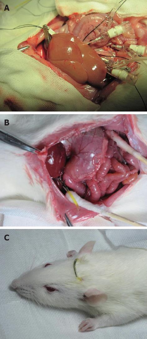
Figure 1 Surgical procedure for a rat model of autologous liver transplantation with continuous external biliary drainage.
A: Lactated Ringer’s solution was perfused by an infusion pump through the portal vein and the abdominal aorta; B: A polyethylene catheter was inserted into the common bile duct for external biliary drainage; C: The free ends of external biliary drainage tube and jejunal fistula tube were connected.
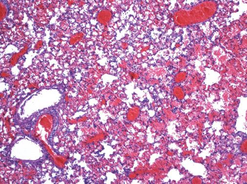
Figure 2 Pathological examination of the lung after the death of three rats in group IV (hematoxylin-eosin; original magnification × 400).
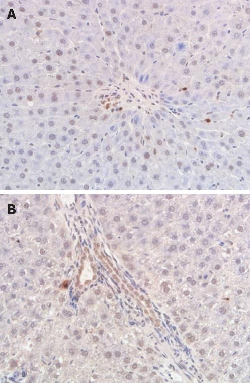
Figure 3 With the prolongation of secondary biliary warm ischemia time, more biliary epithelial cells became apoptotic.
A: Biliary epithelial cell apoptosis in group I at 24 h after hepatic arterial (HA) reperfusion; B: Biliary epithelial cell apoptosis in group IV at 24 h after HA reperfusion (hematoxylin-eosin; original magnification × 400).
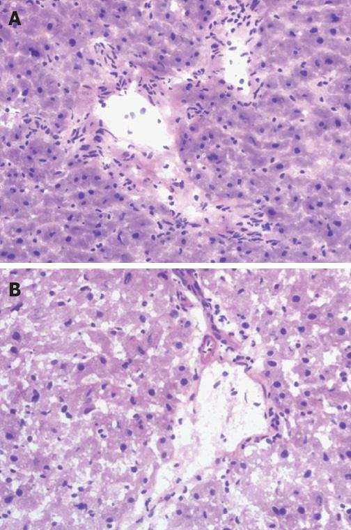
Figure 4 Histological examination of the liver at 24 h after hepatic arterial reperfusion.
A: Cholangiocyte injury can be found in group I; B: More marked injury occurs in group IV (hematoxylin-eosin; original magnification × 400).
- Citation: Zhu XH, Pan JP, Wu YF, Ding YT. Establishment of a rat liver transplantation model with prolonged biliary warm ischemia time. World J Gastroenterol 2012; 18(48): 7194-7200
- URL: https://www.wjgnet.com/1007-9327/full/v18/i48/7194.htm
- DOI: https://dx.doi.org/10.3748/wjg.v18.i48.7194


