Copyright
©2012 Baishideng Publishing Group Co.
World J Gastroenterol. Dec 28, 2012; 18(48): 7184-7193
Published online Dec 28, 2012. doi: 10.3748/wjg.v18.i48.7184
Published online Dec 28, 2012. doi: 10.3748/wjg.v18.i48.7184
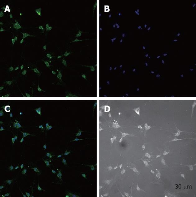
Figure 1 Identification of cultured interstitial cells of Cajal.
A-C: Prolonging the culture to 4-7 d, the cultured interstitial cells of Cajal (ICC), which are identified by c-Kit immunofluorescence, had distinctive shapes such as spindle, triangular or stellar-like with two to five long processes. ICC were fixed with acetone and identified immunologically using a monoclonal c-Kit antibody and Alexa Fluor 488-conjugated secondary fluorescent antibody. Nuclei were stained with Hoechst 33258 dye (B, blue); C: A merged image of A and B; D: A light microscopic image of ICC.
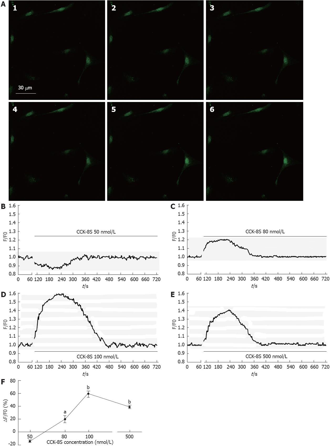
Figure 2 The regulation of sulfated cholecystokinin-8 on [Ca2+]i in cultured interstitial cells of Cajal from the murine gastric antrum.
A1: Fluorescent intensity image of Fluo-3/AM loaded cultured interstitial cells of Cajal (ICC) under normal conditions; A2-6: Fluorescent intensity gradually increased in the presence of cholecystokinin-8 (CCK-8S) (100 nmol/L); B-E: Effects of different concentrations of sulfated CCK-8S on mean [Ca2+]i. In each case, cells from at least five different cell cultures; F: Effects of CCK-8S were estimated as percentage of ΔF/F0, where F0 was derived from the averaged intensity of the first 10-30 frames minus the background in the cell-free region and ΔF is fluorescent intensity of the response minus F0. aP < 0.05, bP < 0.01 vs control.
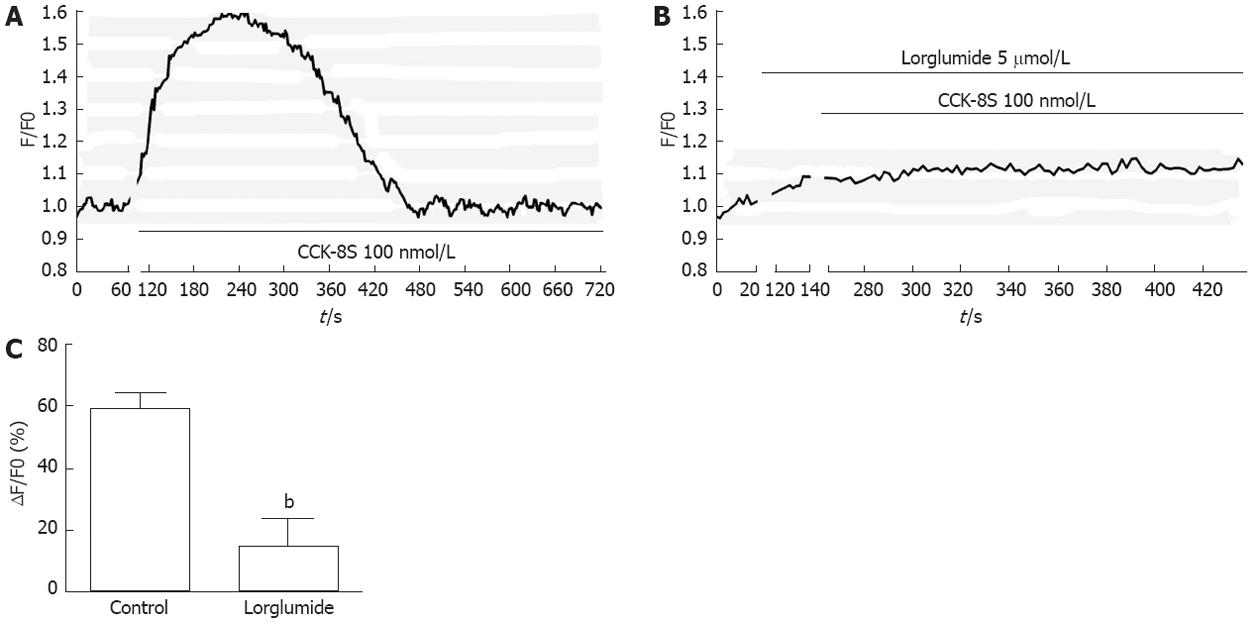
Figure 3 Sulfated cholecystokinin-8 activates interstitial cells of Cajal through the cholecystokinin1 receptor.
A: Compared with the control; B: Lorglumide significantly inhibited cholecystokinin-8 (CCK-8S)-induced increase in [Ca2+]i of interstitial cells of Cajal; C: Quantification of [Ca2+]i changes shown in A and B. Each experiment was repeated at least three times. bP < 0.01 vs control.
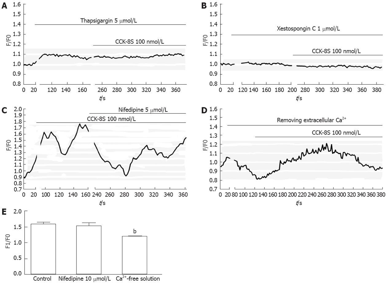
Figure 4 Sulfated cholecystokinin-8 induced calcium mobilization in cultured interstitial cells of Cajal.
A, B: Pretreatment with 5 μmol/L thapsigargin (A) or 1 μmol/L xestospongin C (B) completely abolished sulfated cholecystokinin-8 (CCK-8S)-induced [Ca2+]i increases; C: Addition of nifedipine resulted in a smaller peak of [Ca2+]i in comparison with normal conditions; D: The CCK-8S-elicited [Ca2+]i increase in the calcium-free medium was lower than that in the calcium-containing buffer; E: Quantification of [Ca2+]i changes following addition of nifedipine or removal of extracellular Ca2+. The fluorescence was normalized as F1/F0 (F1: Maximal fluorescence after drug addition; F0: Basal fluorescence before drug addition). bP < 0.01 vs control.
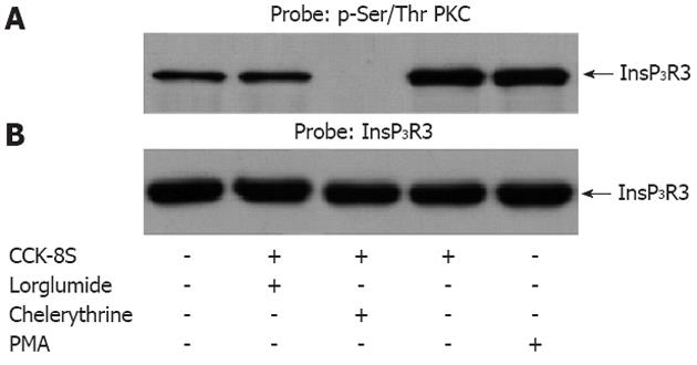
Figure 5 Sulfated cholecystokinin-8 stimulation of interstitial cells of Cajal resulted in the protein kinase C-dependent phosphorylation of type III inositol 1,4,5-triphosphate receptor.
A: Western blots of proteins were immunoprecipitated with type III inositol 1,4,5-triphosphate receptor (InsP3R3)-specific antibody. The immunoprecipitated proteins were probed with antibody specific for phosphorylated Ser/Thr protein kinase C (PKC) substrate sequences. The sulfated cholecystokinin-8 (CCK-8S)-induced phosphorylation of InsP3R3 was apparently inhibited by pretreatment with chelerythine. Pretreatment with lorglumide (5 μmol/L) could significantly reduce the CCK-8S intensified phosphorylation of InsP3R3. In the positive control group, treatment of cells with phorbol-12-myristate-13-acetate (PMA) also resulted in an enhanced phosphorylation of InsP3R3; B: The nitrocellulose membrane in A was stripped and reprobed with InsP3R3 (1:1000) to determine the levels of InsP3R3 immunoprecipitated. Each cell sample contained nearly equal amounts of InsP3R3.
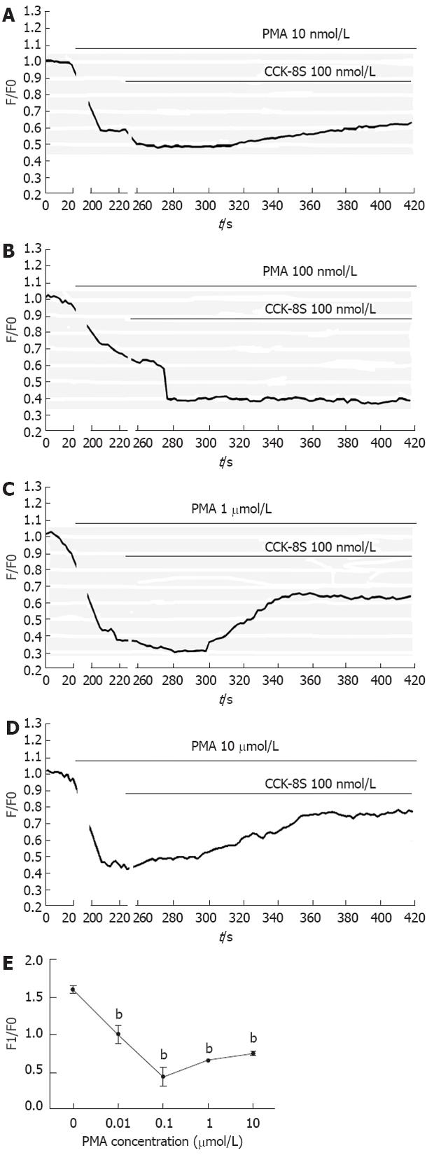
Figure 6 Effects of phorbol-12-myristate-13-acetate on sulfated cholecystokinin-8-evoked Ca2+ signaling in interstitial cells of Cajal.
Fluo-3-loaded interstitial cells of Cajal were pretreated with various concentrations of Phorbol-12-myristate-13-acetate (PMA) (A: 10 nmol/L; B: 100 nmol/L; C: 1 μmol/L; D: 10 μmol/L) for 4 min before addition of sulfated cholecystokinin-8 (CCK-8S) (100 nmol/L). All could significantly reduce the CCK-8S-evoked [Ca2+]i response; E: Quantification of [Ca2+]i changes shown in A-D. Each experiment was repeated at least three times. bP < 0.01 vs control.
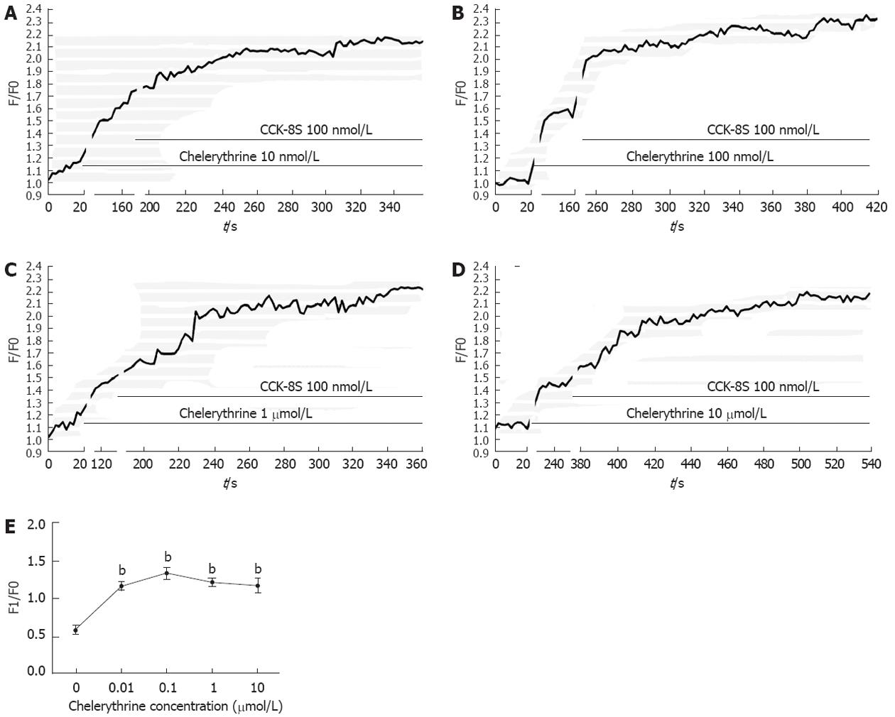
Figure 7 Effects of chelerythrine on Sulfated cholecystokinin-8-evoked Ca2+ signaling in interstitial cells of Cajal.
Fluo-3-loaded interstitial cells of Cajal were pretreated with various concentrations of chelerythrine (A: 10 nmol/L; B: 100 nmol/L; C: 1 μmol/L; and D: 10 μmol/L) for 2 min before administration of cholecystokinin-8 (CCK-8S) (100 nmol/L). All could significantly increase the CCK-8S-evoked [Ca2+]i response; E: Quantification of [Ca2+]i changes shown in A-D. Each experiment was repeated at least three times. bP < 0.01 vs control.
- Citation: Gong YY, Si XM, Lin L, Lu J. Mechanisms of cholecystokinin-induced calcium mobilization in gastric antral interstitial cells of Cajal. World J Gastroenterol 2012; 18(48): 7184-7193
- URL: https://www.wjgnet.com/1007-9327/full/v18/i48/7184.htm
- DOI: https://dx.doi.org/10.3748/wjg.v18.i48.7184


