Copyright
©2012 Baishideng Publishing Group Co.
World J Gastroenterol. Dec 28, 2012; 18(48): 7184-7193
Published online Dec 28, 2012. doi: 10.3748/wjg.v18.i48.7184
Published online Dec 28, 2012. doi: 10.3748/wjg.v18.i48.7184
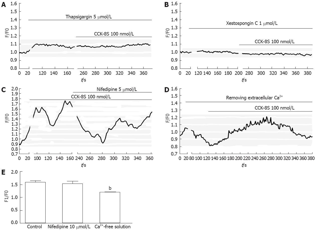
Figure 4 Sulfated cholecystokinin-8 induced calcium mobilization in cultured interstitial cells of Cajal.
A, B: Pretreatment with 5 μmol/L thapsigargin (A) or 1 μmol/L xestospongin C (B) completely abolished sulfated cholecystokinin-8 (CCK-8S)-induced [Ca2+]i increases; C: Addition of nifedipine resulted in a smaller peak of [Ca2+]i in comparison with normal conditions; D: The CCK-8S-elicited [Ca2+]i increase in the calcium-free medium was lower than that in the calcium-containing buffer; E: Quantification of [Ca2+]i changes following addition of nifedipine or removal of extracellular Ca2+. The fluorescence was normalized as F1/F0 (F1: Maximal fluorescence after drug addition; F0: Basal fluorescence before drug addition). bP < 0.01 vs control.
- Citation: Gong YY, Si XM, Lin L, Lu J. Mechanisms of cholecystokinin-induced calcium mobilization in gastric antral interstitial cells of Cajal. World J Gastroenterol 2012; 18(48): 7184-7193
- URL: https://www.wjgnet.com/1007-9327/full/v18/i48/7184.htm
- DOI: https://dx.doi.org/10.3748/wjg.v18.i48.7184


