Copyright
©2012 Baishideng Publishing Group Co.
World J Gastroenterol. Dec 21, 2012; 18(47): 6974-6980
Published online Dec 21, 2012. doi: 10.3748/wjg.v18.i47.6974
Published online Dec 21, 2012. doi: 10.3748/wjg.v18.i47.6974
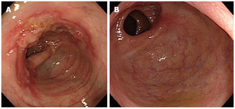
Figure 1 Typical colonoscopic views of intestinal tuberculosis.
A: Colonoscopy shows a circumferential ulcer with edematous flared nodules in the ascending colon (patient 1); B: Colonoscopy shows a whitish mucosal area with an absence of the normal vascular pattern of healed ulcer scars in the ascending colon. Note the concomitant active ulcers in the proximal colon (patient 2).
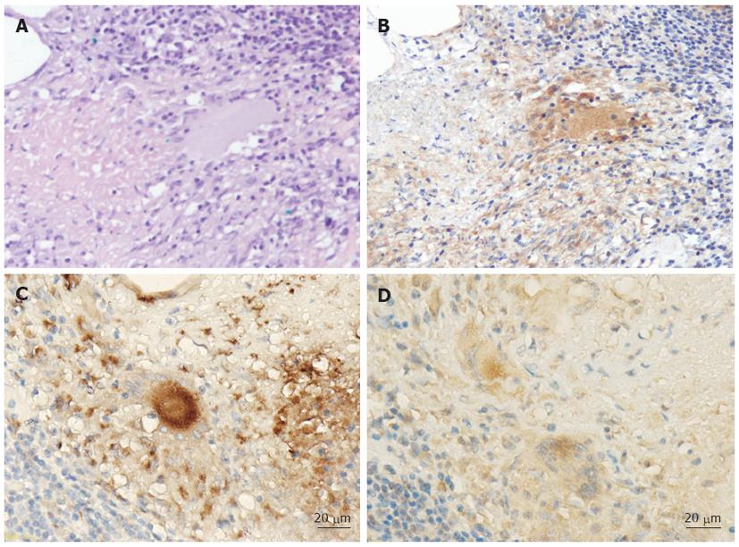
Figure 2 Histopathological views of tuberculous granuloma and localization of the mycobacterial antigen in the colonic lesion (patient 8).
A: A granuloma, surrounded by inflammatory lymphocytes, is present in the lamina propria (hematoxylin and eosin, × 200); B: Immunohistochemical staining view of the colonic specimen (× 200). Note that the mycobacterial antigens (brownish granular matter) are present in the cytoplasm of epithelioid histiocytes and multinucleated giant cells in the granuloma; C: Cells stained with the pan-macrophage marker CD68 antibody are present in the granuloma (× 400); D: The mycobacterial antigens (brownish granular matter) are present in the cytoplasm of the macrophages in the granuloma (× 400).
-
Citation: Ihama Y, Hokama A, Hibiya K, Kishimoto K, Nakamoto M, Hirata T, Kinjo N, Cash HL, Higa F, Tateyama M, Kinjo F, Fujita J. Diagnosis of intestinal tuberculosis using a monoclonal antibody to
Mycobacterium tuberculosis . World J Gastroenterol 2012; 18(47): 6974-6980 - URL: https://www.wjgnet.com/1007-9327/full/v18/i47/6974.htm
- DOI: https://dx.doi.org/10.3748/wjg.v18.i47.6974


