Copyright
©2012 Baishideng Publishing Group Co.
World J Gastroenterol. Aug 21, 2012; 18(31): 4136-4144
Published online Aug 21, 2012. doi: 10.3748/wjg.v18.i31.4136
Published online Aug 21, 2012. doi: 10.3748/wjg.v18.i31.4136
Figure 1 Two-dimensional double contrast-enhanced ultrasound imaging.
A: The picture showing intravenous contrast harmonic imaging with the echo-free gastric cavity and three layers of normal gastric wall (arrow); B: The picture showing oral contrast imaging of normal gastric wall (arrow) and cavity filled with echogenic contrast agent.
Figure 2 Double contrast-enhanced ultrasound imaging of gastric ulceration.
A: Three-dimensional (3D) double contrast-enhanced ultrasound imaging showed the gastric cavity and wall with a focal defect area consistent with an ulcer (arrow); B: Another 3D imaging with different angle showed the ulcerative lesion (arrow) and the folds of gastric wall with pseudo-color which similar to gastroscopic imaging; C: The ulcerative lesion (arrow) is seen on gastroscope imaging.
Figure 3 Double contrast-enhanced ultrasound imaging of gastric polyp.
A: Two-dimensional double contrast-enhanced ultrasound (DCUS) imaging displayed a polyp with a wide base projecting into the gastric cavity. Contrast enhancement was seen on both polyp and gastric wall; B: Three-dimensional DCUS imaging of the polyp showed in figure A; C: The surgical specimen of the polyp confirmed the DCUS finding.
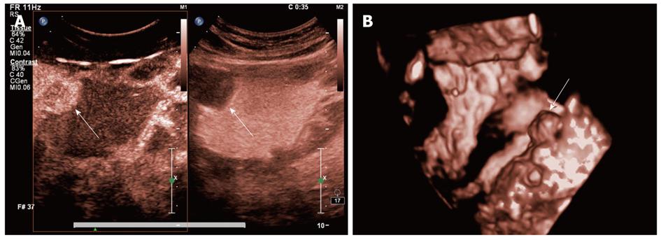
Figure 4 Double contrast-enhanced ultrasound imaging of gastric stromal tumor.
A: Two-dimensional double contrast-enhanced ultrasound (DCUS) images displayed a anechoic mass into the gastric cavity in oral contrast ultrasonography (right figure), from which we hardly judged whether it was cystic or solid lesion; but the intravenous contrast imaging (left figure) showed there was contrast agent enhancement in the focus of infection (arrow); B: Three-dimensional DCUS imaging displayed the tumor elevated to the gastric cavity (arrow).
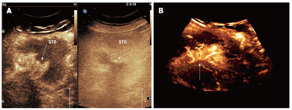
Figure 5 Double contrast-enhanced ultrasound imaging of ulcerative gastric cancer.
A: Two-dimensional double contrast-enhanced ultrasound (DCUS) images (conventional imaging on the right and harmonic imaging on the left) showed a contrast-enhanced mass with crater-like ulcerative defect (arrow); B: Three-dimensional DCUS imaging showed distorted nourishing vasculature within the gastric cancer (arrow).
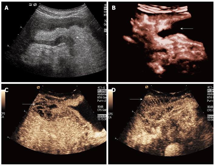
Figure 6 Double contrast-enhanced ultrasound imaging of infiltrative gastric cancer.
A: An oral contrast image showed diffusely thickened gastric wall with narrowed gastric cavity (arrow) in a patient with gastric cancer; B: Three-dimensional double contrast-enhanced ultrasound (DCUS) imaging showed thickened gastric wall and narrowed echo-free gastric cavity (arrow); C, D: Two-dimensional DCUS images showed fence-like tumor vessels penetrating through thickened gastric wall in the arterial phase (arrows).
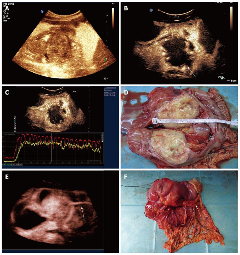
Figure 7 Double contrast-enhanced ultrasound imaging of mass-type gastric cancer.
A: Oral contrast imaging revealed a tumor (arrow) extended into the back wall of stomach, which could not be seen by gastroscopy; B: Two-dimensional double contrast-enhanced ultrasound (DCUS) imaging showed the tumor attached to the posterior wall and enhanced in peripheral areas with the center of non-enhancement at early arterial phase; C: A contrast time-intensity curve of the lesion showed that the arrival time was shorter and the peak intensity was higher in the peripheral lesion (red curve) than those in normal gastric wall (yellow curve); D: The surgical specimen demonstrated the tumor with central necrosis which consistent to DCUS findings; E: Three-dimensional DCUS imaging showed the outer-growing lesion with a tumor feeding vessel (arrow); F: The feeding vessel on the gross specimen was seen and coincident with the DCUS imaging.
- Citation: Shi H, Yu XH, Guo XZ, Guo Y, Zhang H, Qian B, Wei ZR, Li L, Wang XC, Kong ZX. Double contrast-enhanced two-dimensional and three-dimensional ultrasonography for evaluation of gastric lesions. World J Gastroenterol 2012; 18(31): 4136-4144
- URL: https://www.wjgnet.com/1007-9327/full/v18/i31/4136.htm
- DOI: https://dx.doi.org/10.3748/wjg.v18.i31.4136


