Copyright
©2012 Baishideng Publishing Group Co.
World J Gastroenterol. Aug 21, 2012; 18(31): 4136-4144
Published online Aug 21, 2012. doi: 10.3748/wjg.v18.i31.4136
Published online Aug 21, 2012. doi: 10.3748/wjg.v18.i31.4136
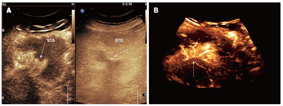
Figure 5 Double contrast-enhanced ultrasound imaging of ulcerative gastric cancer.
A: Two-dimensional double contrast-enhanced ultrasound (DCUS) images (conventional imaging on the right and harmonic imaging on the left) showed a contrast-enhanced mass with crater-like ulcerative defect (arrow); B: Three-dimensional DCUS imaging showed distorted nourishing vasculature within the gastric cancer (arrow).
- Citation: Shi H, Yu XH, Guo XZ, Guo Y, Zhang H, Qian B, Wei ZR, Li L, Wang XC, Kong ZX. Double contrast-enhanced two-dimensional and three-dimensional ultrasonography for evaluation of gastric lesions. World J Gastroenterol 2012; 18(31): 4136-4144
- URL: https://www.wjgnet.com/1007-9327/full/v18/i31/4136.htm
- DOI: https://dx.doi.org/10.3748/wjg.v18.i31.4136


