Copyright
©2012 Baishideng Publishing Group Co.
World J Gastroenterol. Aug 21, 2012; 18(31): 4136-4144
Published online Aug 21, 2012. doi: 10.3748/wjg.v18.i31.4136
Published online Aug 21, 2012. doi: 10.3748/wjg.v18.i31.4136
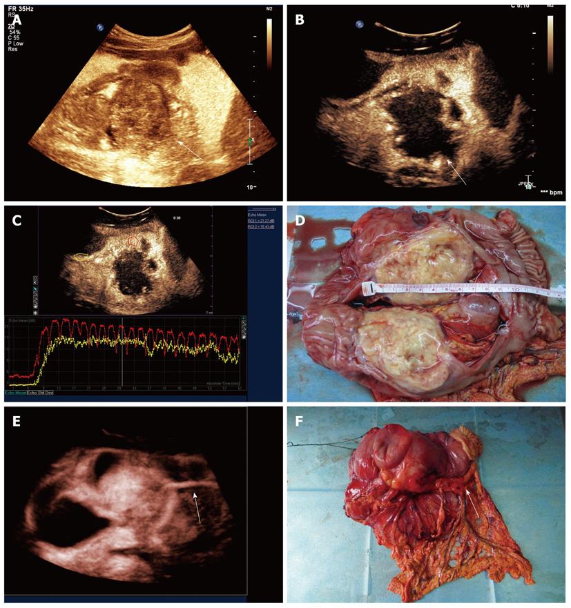
Figure 7 Double contrast-enhanced ultrasound imaging of mass-type gastric cancer.
A: Oral contrast imaging revealed a tumor (arrow) extended into the back wall of stomach, which could not be seen by gastroscopy; B: Two-dimensional double contrast-enhanced ultrasound (DCUS) imaging showed the tumor attached to the posterior wall and enhanced in peripheral areas with the center of non-enhancement at early arterial phase; C: A contrast time-intensity curve of the lesion showed that the arrival time was shorter and the peak intensity was higher in the peripheral lesion (red curve) than those in normal gastric wall (yellow curve); D: The surgical specimen demonstrated the tumor with central necrosis which consistent to DCUS findings; E: Three-dimensional DCUS imaging showed the outer-growing lesion with a tumor feeding vessel (arrow); F: The feeding vessel on the gross specimen was seen and coincident with the DCUS imaging.
- Citation: Shi H, Yu XH, Guo XZ, Guo Y, Zhang H, Qian B, Wei ZR, Li L, Wang XC, Kong ZX. Double contrast-enhanced two-dimensional and three-dimensional ultrasonography for evaluation of gastric lesions. World J Gastroenterol 2012; 18(31): 4136-4144
- URL: https://www.wjgnet.com/1007-9327/full/v18/i31/4136.htm
- DOI: https://dx.doi.org/10.3748/wjg.v18.i31.4136


