Copyright
©2012 Baishideng Publishing Group Co.
World J Gastroenterol. Jun 21, 2012; 18(23): 2956-2965
Published online Jun 21, 2012. doi: 10.3748/wjg.v18.i23.2956
Published online Jun 21, 2012. doi: 10.3748/wjg.v18.i23.2956
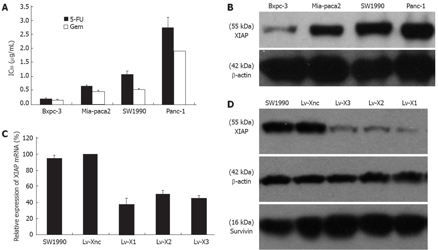
Figure 1 X-linked inhibitor of apoptosis protein expression analysis and selection of the RNAi target for X-linked inhibitor of apoptosis protein.
A, B: The X-linked inhibitor of apoptosis protein (XIAP) protein level and IC50 for Panc-1, SW1990, Mia-paca2 and Bxpc-3 cell lines. R2 = 0.87 (Spearman correlation) between IC50 and relative XIAP expression, P < 0.05; C: Relative expression of XIAP mRNA after transfection and selection with puromycin; D: Downregulatory effect of lentivirus-mediated RNAi on XIAP in SW1990 cells. Lv-X1, Lv-X2, Lv-X3 efficiently suppressed XIAP expression. The results also showed that survivin was not affected by any lentivirus. Lv-Xnc, which was transfected with the non-sense lentivirus vector, was set as the calibrator with the relative expression value of “1”. β-actin was used as the internal loading control in three independent experiments. 5-FU: 5-fluorouracil.
Figure 2 Lentivirus-mediated shRNA targeting the X-linked inhibitor of apoptosis protein gene inhibits the growth of human pancreatic cancer SW1990 cells in vitro and in vivo and promotes chemosensitivity to drugs.
A: Lv-X1, Lv-X2, Lv-X3 promoted 5-fluorouracil (5-FU) or gemcitabine-induced cytotoxicity. The IC50 was determined by 3-(4,5-cimethylthiazol-2-yl)-2,5-diphenyl tetrazolium bromide assay after exposure to 5-FU or gemcitabine for 72 h; B: The growth curves of different stably transfected cell lines; C: Results of colony formation assay. Lv-X1 had much fewer colonies than Lv-Xnc and untransfected cells. These experiments were performed 3 times; D: SW1990, Lv-Xnc and Lv-X1 cells growth in BALB/c nude mice (P < 0.05, from day 12 to day 28).
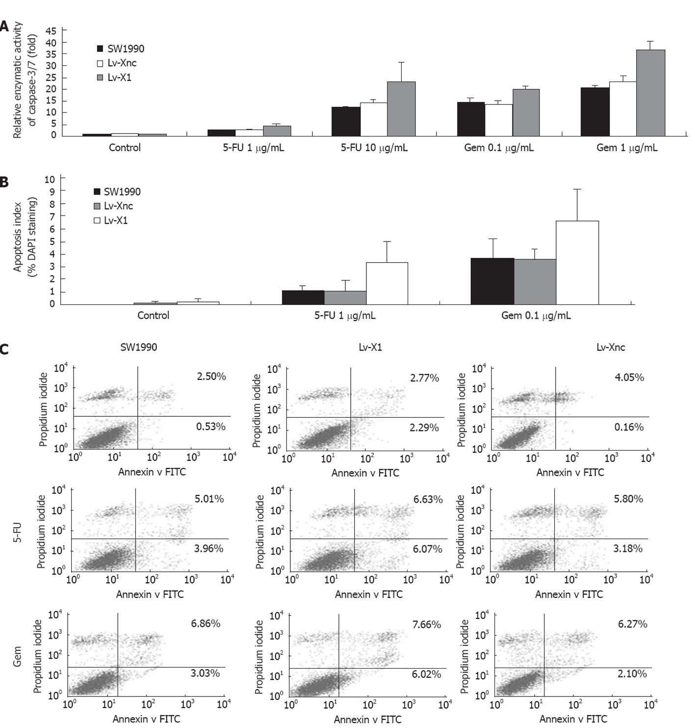
Figure 3 Apoptosis analysis.
A: Relative enzymatic activity of caspase-3/7; B: Apoptosis examined by 4’,6-diamidino-2-phenylindole (DAPI) staining (200×), apoptosis index (%) = Apoptotic cells/Total cells × 100% (Seen in a same microscopic field and 3 randomized microscopic fields chosen for counting) using DAPI staining. Either gemcitabine or 5-fluorouracil (5-FU) kills cancer cells by inducing apoptosis, and X-linked inhibitor of apoptosis protein knocked out by Lv-X1 can enhance the capacity of the 2 drugs; C: Flow cytometry: The apoptotic rate of Lv-X1 and Lv-Xnc slightly increased but there was no significant difference among SW1990, Lv-X1 and Lv-Xnc. While the apoptotic rate of Lv-X1 + 5-FU and Lv-X1 + gemcitabine increased (12.7% and 13.68%, respectively, P < 0.05). FITC: Fluorescein isothiocyanate.
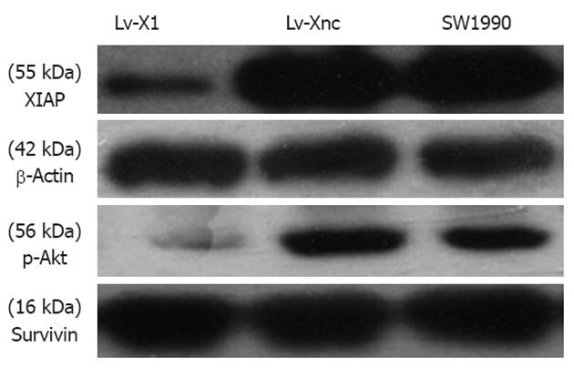
Figure 4 Immunoblot analysis for p-Akt in SW1990 cells after stable transfection.
Downregulation of X-linked inhibitor of apoptosis protein with Lv-X1 largely decreased p-Akt protein levels and survivin was not affected (6 mo in culture).
- Citation: Jiang C, Yi XP, Shen H, Li YX. Targeting X-linked inhibitor of apoptosis protein inhibits pancreatic cancer cell growth through p-Akt depletion. World J Gastroenterol 2012; 18(23): 2956-2965
- URL: https://www.wjgnet.com/1007-9327/full/v18/i23/2956.htm
- DOI: https://dx.doi.org/10.3748/wjg.v18.i23.2956


