Copyright
©2012 Baishideng Publishing Group Co.
World J Gastroenterol. Jun 21, 2012; 18(23): 2956-2965
Published online Jun 21, 2012. doi: 10.3748/wjg.v18.i23.2956
Published online Jun 21, 2012. doi: 10.3748/wjg.v18.i23.2956
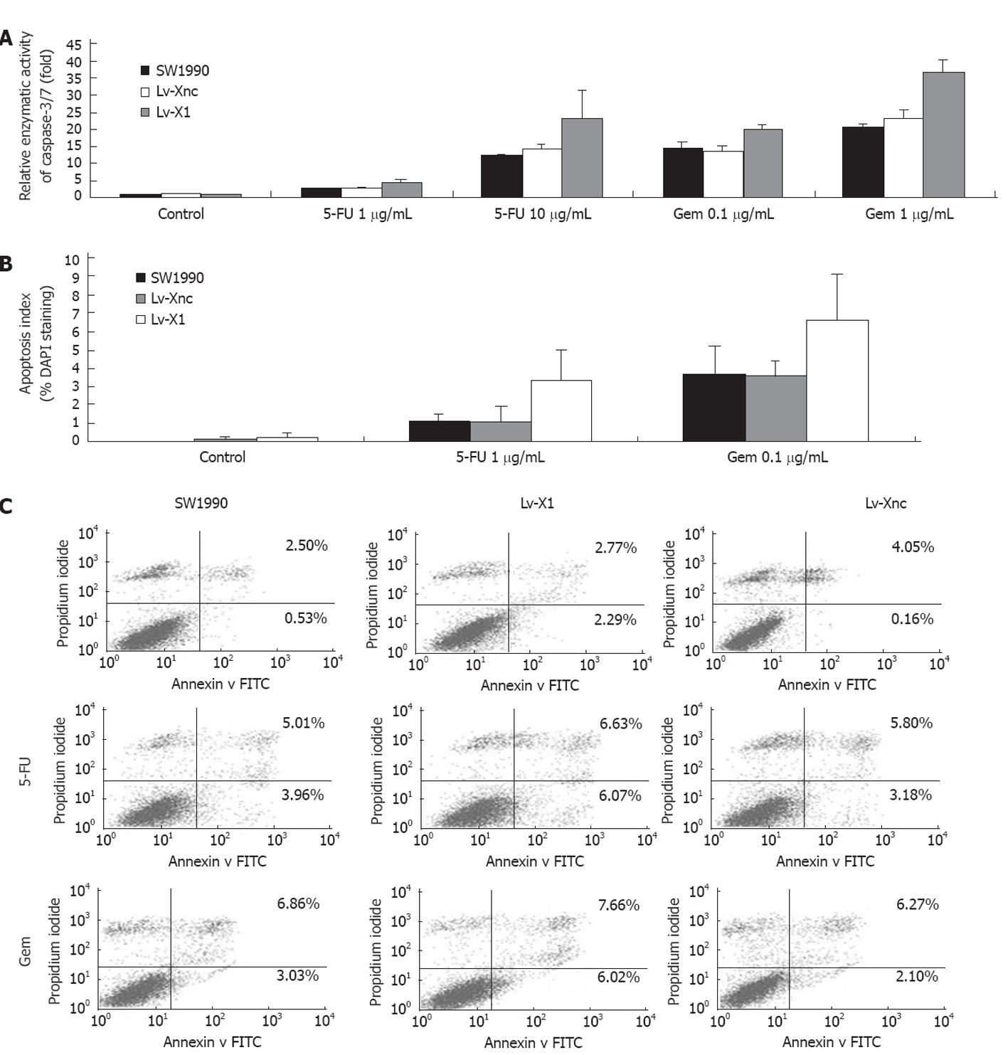
Figure 3 Apoptosis analysis.
A: Relative enzymatic activity of caspase-3/7; B: Apoptosis examined by 4’,6-diamidino-2-phenylindole (DAPI) staining (200×), apoptosis index (%) = Apoptotic cells/Total cells × 100% (Seen in a same microscopic field and 3 randomized microscopic fields chosen for counting) using DAPI staining. Either gemcitabine or 5-fluorouracil (5-FU) kills cancer cells by inducing apoptosis, and X-linked inhibitor of apoptosis protein knocked out by Lv-X1 can enhance the capacity of the 2 drugs; C: Flow cytometry: The apoptotic rate of Lv-X1 and Lv-Xnc slightly increased but there was no significant difference among SW1990, Lv-X1 and Lv-Xnc. While the apoptotic rate of Lv-X1 + 5-FU and Lv-X1 + gemcitabine increased (12.7% and 13.68%, respectively, P < 0.05). FITC: Fluorescein isothiocyanate.
- Citation: Jiang C, Yi XP, Shen H, Li YX. Targeting X-linked inhibitor of apoptosis protein inhibits pancreatic cancer cell growth through p-Akt depletion. World J Gastroenterol 2012; 18(23): 2956-2965
- URL: https://www.wjgnet.com/1007-9327/full/v18/i23/2956.htm
- DOI: https://dx.doi.org/10.3748/wjg.v18.i23.2956


