Copyright
©2012 Baishideng Publishing Group Co.
World J Gastroenterol. Jun 7, 2012; 18(21): 2727-2729
Published online Jun 7, 2012. doi: 10.3748/wjg.v18.i21.2727
Published online Jun 7, 2012. doi: 10.3748/wjg.v18.i21.2727
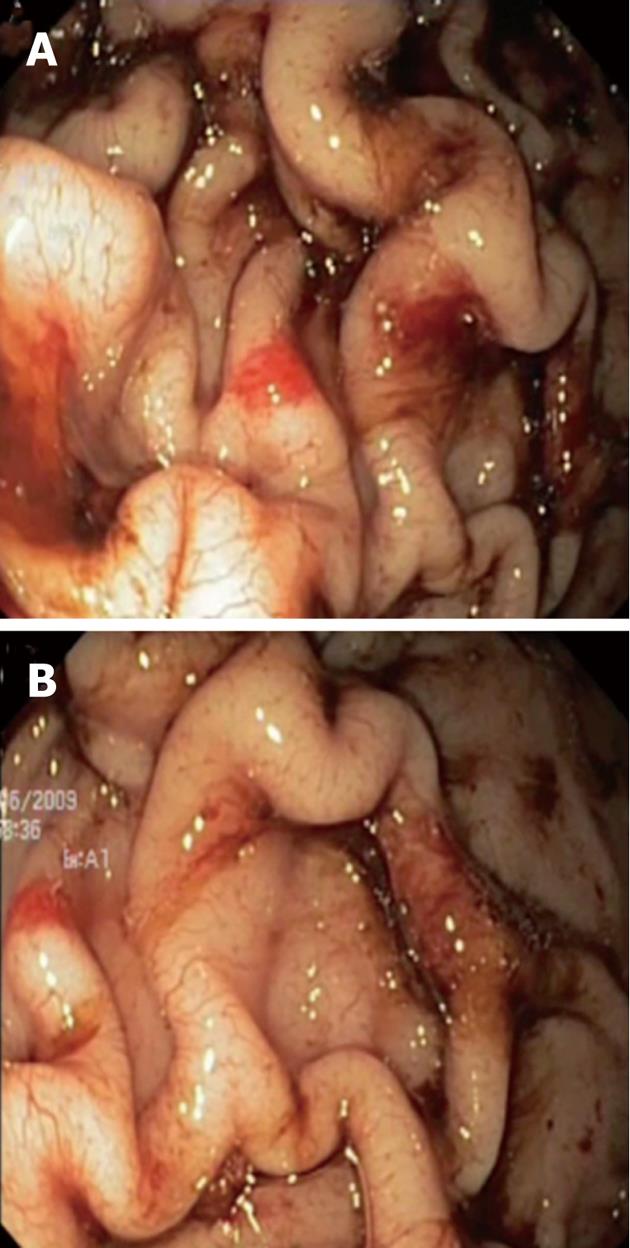
Figure 1 Endoscopic view of markedly thickened gastric folds, with overlying erosions and exudates involving fundus (A) and corpus (B).
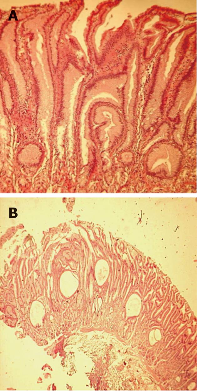
Figure 2 Histopathology from endoscopic mucosal resection (HE staining; A: 40×, B: 20×) shows elongated, tortuous and cystically dilated foveolar glands, discontinuous atrophy of gastric glands and significant reduction of parietal cells.
- Citation: Nardo GD, Oliva S, Aloi M, Ferrari F, Frediani S, Marcheggiano A, Cucchiara S. A pediatric non-protein losing Menetrier's disease successfully treated with octreotide long acting release. World J Gastroenterol 2012; 18(21): 2727-2729
- URL: https://www.wjgnet.com/1007-9327/full/v18/i21/2727.htm
- DOI: https://dx.doi.org/10.3748/wjg.v18.i21.2727


