Copyright
©2012 Baishideng Publishing Group Co.
World J Gastroenterol. May 7, 2012; 18(17): 2035-2042
Published online May 7, 2012. doi: 10.3748/wjg.v18.i17.2035
Published online May 7, 2012. doi: 10.3748/wjg.v18.i17.2035
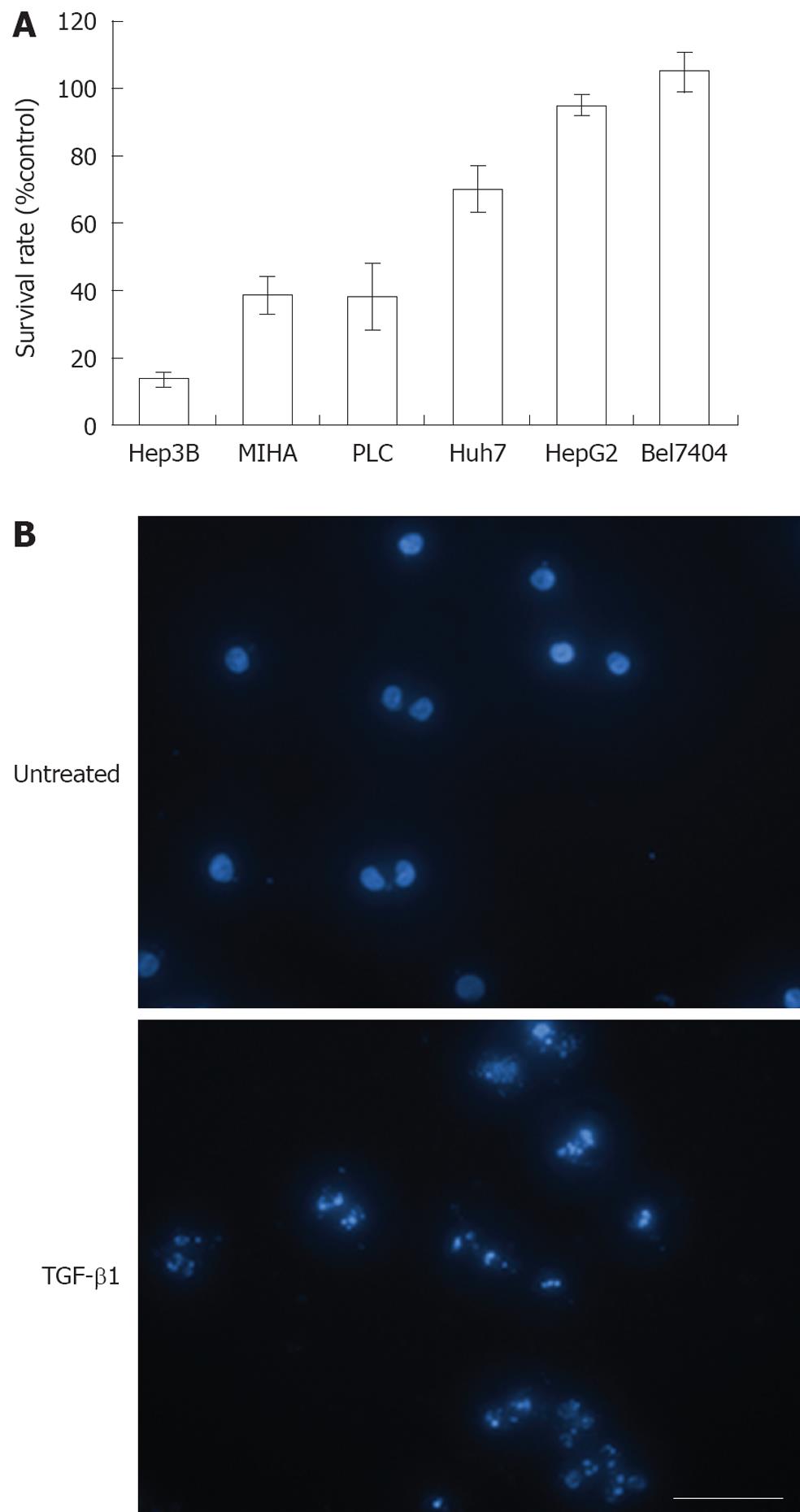
Figure 1 Susceptibilities of various human hepatocyte (MIHA) and hepatocellular carcinoma cells (Hep3B, PLC, Huh7, HepG2, and Bel7404) to transforming growth factor-β.
A: Cells were treated with 5 ng/mL transforming growth factor (TGF)-β1 for 96 h and cell survival was then determined by methylthiazoletetrazolium assays; B: Nuclear morphology of apoptotic TGF-β-treated Hep3B cells demonstrated by 4’,6-Diamidino-2-phenylindole staining and examined by fluorescence microscopy. Scale bar: 50 μm, 400 ×.
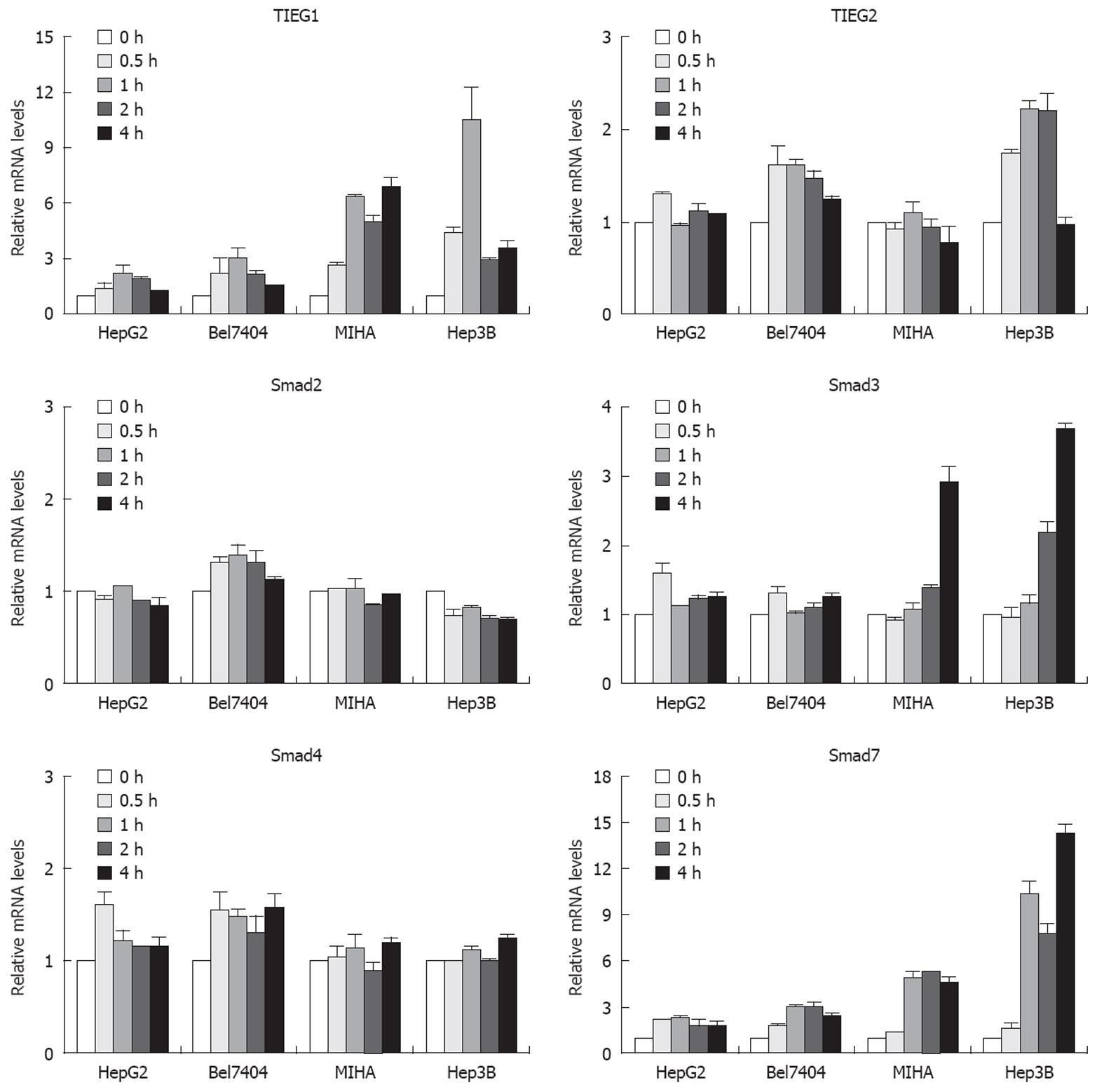
Figure 2 Differential responses of TIEGs and Smads in various TGF-β1-treated cells.
Cells were treated with 5 ng/mL TGF-β1 for up to 4 h. At indicated time intervals, the mRNA levels of TIEG1, TIEG2, Smad2, Smad3, Smad4, and Smad7 were determined by quantitative real-time RT-PCRs. Data were presented as mRNA levels relative to that of time 0 from three independent experiments.
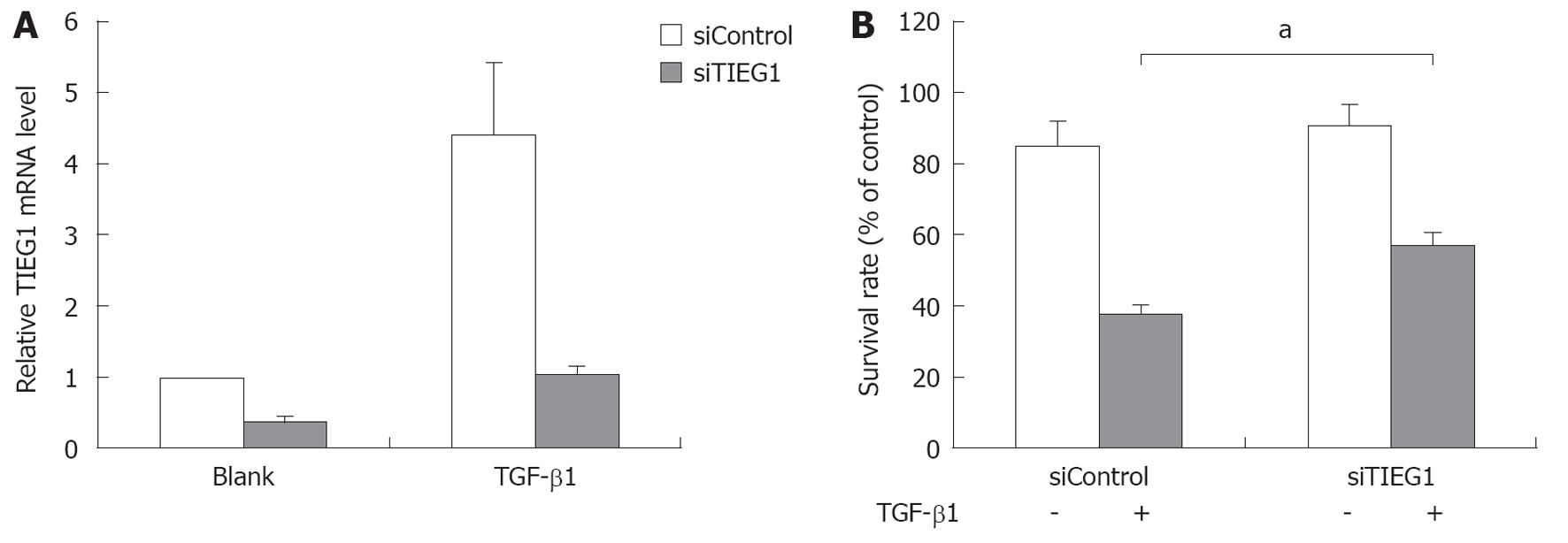
Figure 3 Downregulation of transforming growth factor-β-inducible early gene 1 by RNA interference (siRNA) decreased the transforming growth factor-β1 susceptibility of Hep3B cells.
A: Decreases in the transforming growth factor (TGF)-β-inducible early gene 1 (TIEG1) mRNA level in siRNAs targeting TIEG1 (siTIEG1)-transfected Hep3B cells with or without TGF-β1 treatment. Cells were transfected with siTIEG1 prior to the TGF-β1 treatments described above. After 4 h of treatment, mRNA levels of TIEG1 were determined by quantitative reverse transcriptase-polymerase chain reaction; B: Increases in the survivability of the siTIEG1-transfected and the TGF-β1-treated Hep3B cells. The decreased growth inhibitory response of Hep3B cells to TGF-β1 treatment was determined by methylthiazoletetrazolium assays after 72 h of TGF-β1 treatment. aP < 0.05.
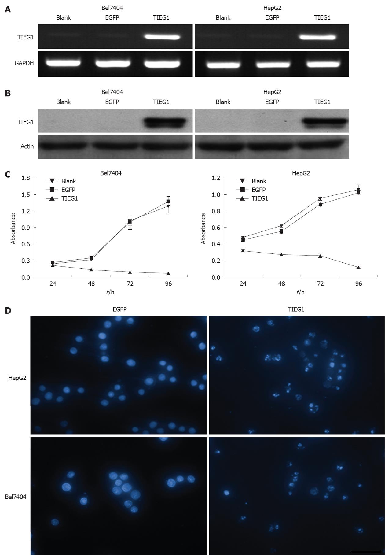
Figure 4 The overexpression of transforming growth factor-β-inducible early gene 1 by lentiviral-mediated transfection-induced growth inhibition and apoptosis in transforming growth factor-β1-resistant hepatoma Bel7404 and HepG2 cells.
A and B: Increases in mRNA and protein levels of transforming growth factor (TGF)-β-inducible early gene 1 (TIEG1) in Bel7404 and HepG2 cells transfected by lenti-TIEG1, respectively, revealed by reverse transcriptase-polymerase chain reaction and Western blotting analysis; C: The overexpression of TIEG1 by lentiviral-mediated transfection-inhibited cell growth in Bel7404 and HepG2 cells. Cell survival was determined by methylthiazoletetrazolium assays; D: Induction of apoptosis in Bel7404 and HepG2 cells after lenti-TIEG1 transfection for 72 h as shown by 4’,6-Diamidino-2-phenylindole staining. Scale bar: 50 μm, 400 ×.
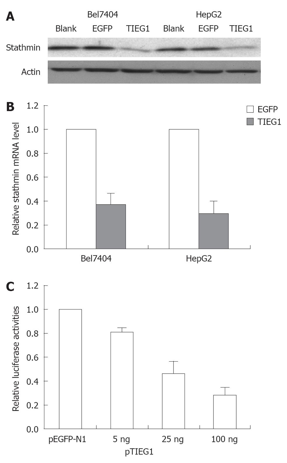
Figure 5 Transcriptional regulation of STMN (stathmin) by transforming growth factor-β-inducible early gene 1.
A and B: Decreases in mRNA and protein levels of STMN in Bel7404 and HepG2 cells transfected by lenti-transforming growth factor-β-inducible early gene 1 (TIEG1), revealed by reverse transcriptase-polymerase chain reaction and Western blotting analyzes; C: Dose-dependent suppression of STMN promoter activity by TIEG1 monitored by a luciferase reporter gene system. Control: pEGFP-N1.
- Citation: Jiang L, Lai YK, Zhang JF, Chan CY, Lu G, Lin MC, He ML, Li JC, Kung HF. Transactivation of the TIEG1 confers growth inhibition of transforming growth factor-β-susceptible hepatocellular carcinoma cells. World J Gastroenterol 2012; 18(17): 2035-2042
- URL: https://www.wjgnet.com/1007-9327/full/v18/i17/2035.htm
- DOI: https://dx.doi.org/10.3748/wjg.v18.i17.2035


