Copyright
©2012 Baishideng Publishing Group Co.
World J Gastroenterol. Apr 28, 2012; 18(16): 1996-1998
Published online Apr 28, 2012. doi: 10.3748/wjg.v18.i16.1996
Published online Apr 28, 2012. doi: 10.3748/wjg.v18.i16.1996
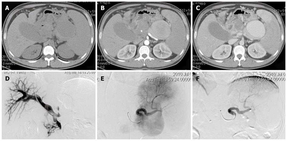
Figure 1 The main foundings of enhanced computed tomography and angiography.
A: An oval cystic low-density lesion located in the area of splenic hilum; B: The lesion was enhanced significantly on the arterial phase; C: Significantly enhanced on the portal phase of contrast-enhanced scan; D: A normal portal pressure and an increased pressure of splenic vein of 23.5 mmHg; E: Splenic arteriovenous fistula manifested by angiography; F: Splenic arteriovenous fistula being totally occluded after embolization.
- Citation: Chen B, Tang CW, Zhang CL, Cao JW, Wei B, Li X. Melena-associated regional portal hypertension caused by splenic arteriovenous fistula. World J Gastroenterol 2012; 18(16): 1996-1998
- URL: https://www.wjgnet.com/1007-9327/full/v18/i16/1996.htm
- DOI: https://dx.doi.org/10.3748/wjg.v18.i16.1996


