Copyright
©2012 Baishideng Publishing Group Co.
World J Gastroenterol. Apr 28, 2012; 18(16): 1892-1902
Published online Apr 28, 2012. doi: 10.3748/wjg.v18.i16.1892
Published online Apr 28, 2012. doi: 10.3748/wjg.v18.i16.1892
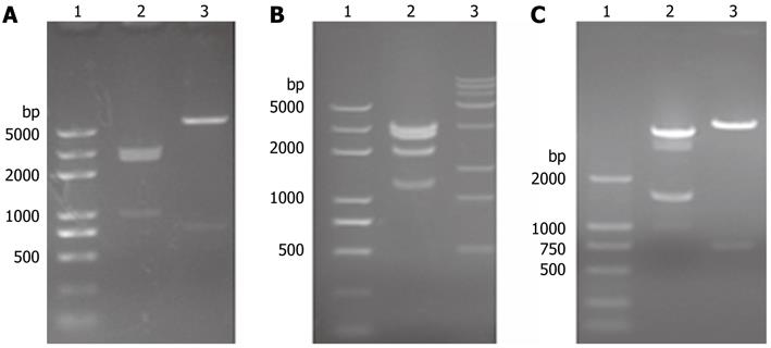
Figure 1 Identification of pTet-on-link, pTet-on-Albumin and pTRE2-urokinase plasminogen activator by restriction endonucleases.
A1 and B1: 2K plus DNA Marker; A2: pTet-on-link/EcoR V + Sal I; A3: pTet-on-link/EcoR V + BamH I; B2: pTet-on-Albumin/BamH I; B3:15K DNA marker; C1: 2K DNA marker; C2: pTRE2-urokinase plasminogen activator (uPA)/pvu II; C3: pTRE2-uPA/Sal I.
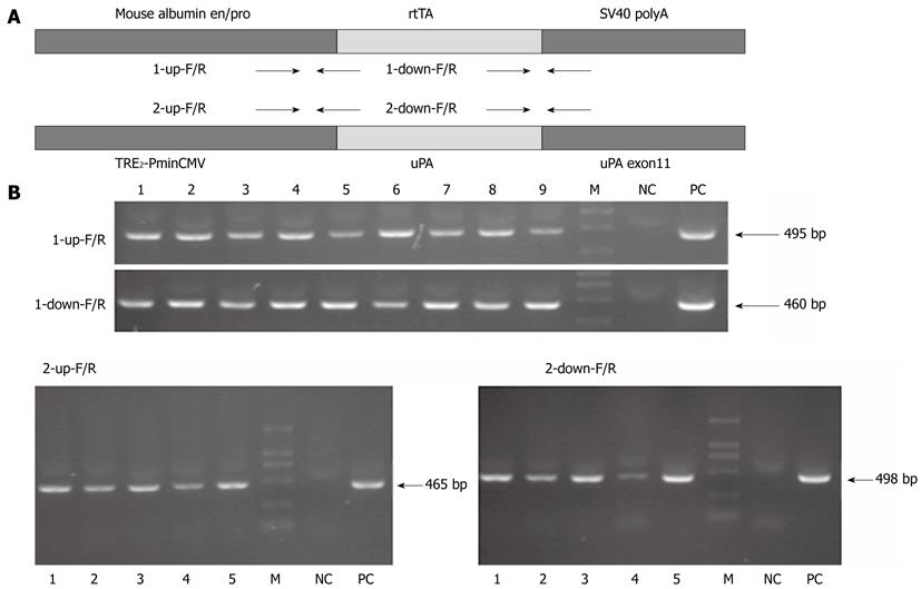
Figure 2 Establishment of albumin-tetracycline reverse transcriptional activator and tetO-urokinase plasminogen activator transgenic mice.
A: The albumin-tetracycline reverse transcriptional activator (rtTA) unit contains the mouse albumin enhancer/promoter, rtTA coding sequence, and SV40 polyA. The tetO-urokinase plasminogen activator (uPA) unit contains the TRE2-PminCMV, uPA cDNA, uPA exon11. Arrowheads depict the positions and directions of the polymerase chain reaction (PCR) primers; B: PCR identification of the transgenic founders. 1-9, PCR identification for the nine albumin-rtTA transgenic founder mice; 1-5, PCR identification for the five tetO-uPA transgenic founder mice. CMV: Cytomegalovirus; M: Marker; NC: Negative control; PC: Positive control.
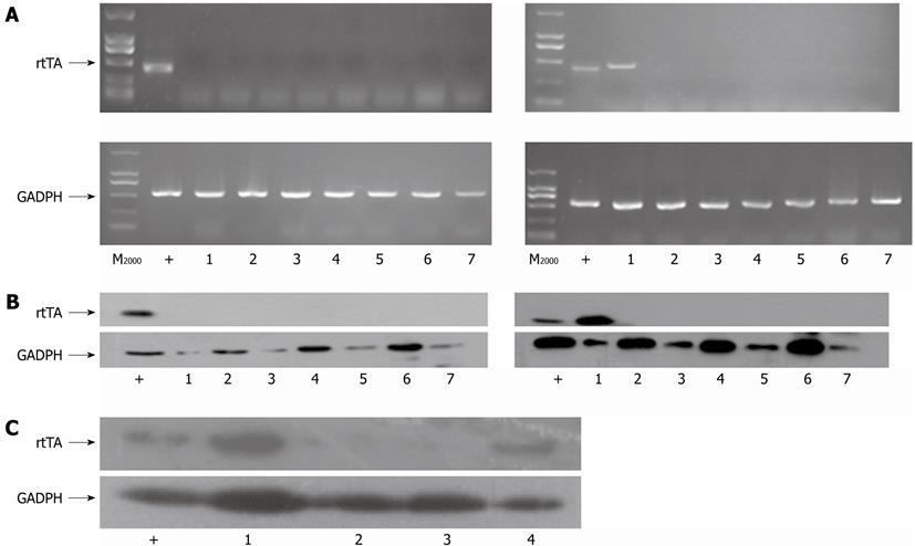
Figure 3 The specific expression of tetracycline reverse transcriptional activator in the livers of albumin-tetracycline reverse transcriptional activatortrangenic mice and in-alb-urokinase plasminogen activator transgenic mice.
A, B: Reverse transcription polymerase chain reaction (A) and Western blotting (B) analysis of tetracycline reverse transcriptional activator (rtTA) and glyceraldehyde-3-phosphate dehydrogenase (GAPDH) expression in different tissues of the 6-8 wk old F1 albumin-rtTA PCR-negative (left for A, B) or positive (right for A, B) transgenic mice. 1: Liver; 2: Brain; 3: Thymus; 4: Heart; 5: Lung; 6: Kidney; 7: Spleen; C: Western blotting analysis of rtTA and GADPH expression in the liver extracts of mice with different genotypes.1: In-alb-urokinase plasminogen activator (uPA) mice group; 2: Liver extracts of wild type mice group; 3: tetO-uPA mice group; 4: Albumin-rtTA mice group. pTet-on transfected Huh7 cell extracts were used as positive control (+).
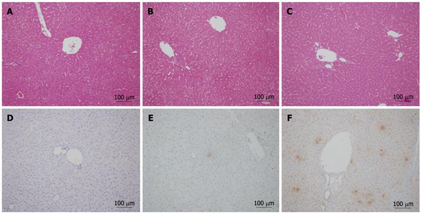
Figure 4 The expression of urokinase plasminogen activator in the livers of in-alb-urokinase plasminogen activator transgenic mice.
A: Histology of livers from wild type (WT) mice; B: Histology of livers from in-alb-urokinase plasminogen activator (uPA) mice without doxycycline (Dox) induction; C: Histology of livers from in-alb-uPA mice with Dox induction; D: uPA expression in hepatocytes from from WT mice; E: uPA expression in hepatocytes from in-alb-uPA mice without Dox induction; F: uPA expression in hepatocytes from in-alb-uPA mice with Dox induction. Magnification, × 20.
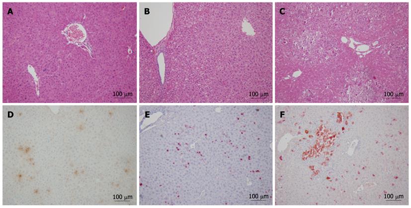
Figure 5 Synergistic injury of liver in in-alb-urokinase plasminogen activator transgenic mice after adeno-associated virus-1.
3hepatitis B virus transfection. Histological and immunohistochemical staining for hepatitis B core antigen in the livers of different groups of mice 20 d later after adeno-associated virus (AAV)-1.3hepatitis B virus (HBV) transfection. A: Histology of the AAV-internal ribosome entry site (IRES) transfected doxycycline (Dox)-induced in-alb-urokinase plasminogen activator (uPA) mice; B: Histology of the AAV-HBV transfected non-induced in-alb-uPA mice; C: Histology of the AAV-HBV transfected Dox-induced in-alb-uPA mice; D: Double-staining immunohistochemical analysis of the AAV-IRES transfected Dox-induced in-alb-uPA mice; E: Double-staining immunohistochemical analysis of the AAV-HBV transfected non-induced in-alb-uPA mice; F: Double-staining immunohistochemical analysis of the AAV-HBV transfected Dox-induced in-alb-uPA mice. Magnification, × 20.
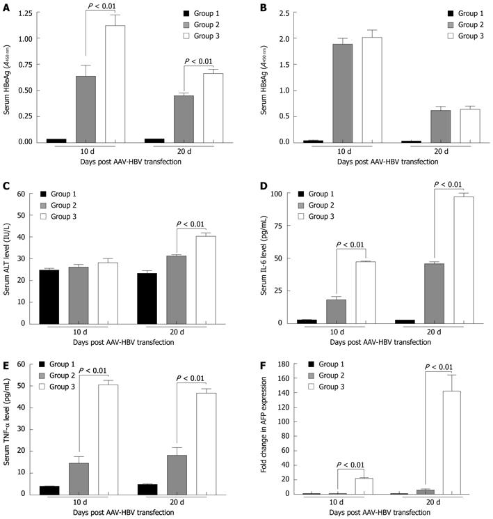
Figure 6 Comparison of the serum hepatitis B e antigen, hepatitis B surface antigen, interleukin-6, tumor necrosis factor-α, alanine aminotransferase levels and hepatic α-fetoprotein mRNA levels between mice from different groups.
Group 1: Adeno-associated virus (AAV)-internal ribosome entry site transfected doxycycline (Dox)-induced in-alb-urokinase plasminogen activator (uPA) mice (n = 6); Group 2: AAV-hepatitis B virus (HBV) transfected non-induced in-alb-uPA mice (n = 6); Group 3: AAV-HBV transfected Dox-induced in-alb-uPA mice (n = 6). HBeAg: Hepatitis B e antigen; HBsAg: Hepatitis B surface antigen; ALT: Alanine aminotransferase; IL: Interleukin; TNF: Tumor necrosis factor; AFP: α-fetoprotein.
- Citation: Zhou XJ, Sun SH, Wang P, Yu H, Hu JY, Shang SC, Zhou YS. Over-expression of uPA increases risk of liver injury in pAAV-HBV transfected mice. World J Gastroenterol 2012; 18(16): 1892-1902
- URL: https://www.wjgnet.com/1007-9327/full/v18/i16/1892.htm
- DOI: https://dx.doi.org/10.3748/wjg.v18.i16.1892


