Copyright
©2012 Baishideng Publishing Group Co.
World J Gastroenterol. Mar 28, 2012; 18(12): 1295-1307
Published online Mar 28, 2012. doi: 10.3748/wjg.v18.i12.1295
Published online Mar 28, 2012. doi: 10.3748/wjg.v18.i12.1295
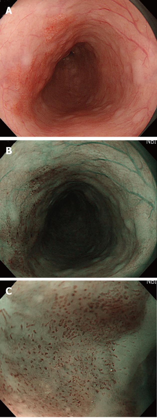
Figure 1 The carcinoma visualized in esophagus.
A: Carcinoma in esophagus is difficult to identify by conventional white light; B: Carcinoma in esophagus can be easily recognized by narrow-band imaging (NBI) as well-demarcated brownish area; C: Intraepithelial papillary capillary loop can be observed by magnifying endoscopy with NBI at the edge of the tumor.
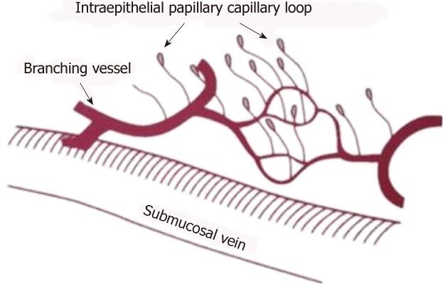
Figure 2 The superficial blood vessels in the squamous esophagus (from Inoue et al[42], with permission, making a little change for the original graph), the intraepithelial papillary capillary loop rises from the branching vessel and terminates in a diffuse.
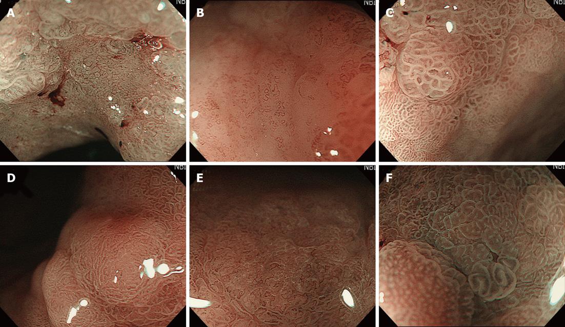
Figure 3 Some typical microvascular and microsurface imaging characteristics visualized in stomach under magnifying endoscopy with narrow band imaging.
A: Fine network pattern, mostly corresponding to well-differentiated adenocarcinoma (0-IIc, gastric); B: Corkscrew pattern, mostly corresponding to the poorly-differentiated adenocarcinoma (0-IIc, gastric); C: Intrastructural irregular vessel, enclosed in villous or papillary fine mucosal structure, had irregular shape characters such as dilation, heterogeneity, abrupt caliber or tortuousness (0-IIb, gastric); D: Regular white opaque substance (WOS), that shows well-organized and symmetrical distribution with a regular reticular pattern and obscures the subepithelial microvascular (MV) pattern (0-IIa adenoma, gastric); E: Irregular WOS, that is present within the cancerous epithelium with an irregular speckled pattern and makes the subepithelial MV pattern cannot be clearly visualized (0-IIa cancer, gastric); F: Light blue crest, defined as a fine, blue-white line on the crests of the epithelial surface in the gastric mucosa may be a distinctive endoscopic finding associated with the presence of histological intestinal metaplasia.
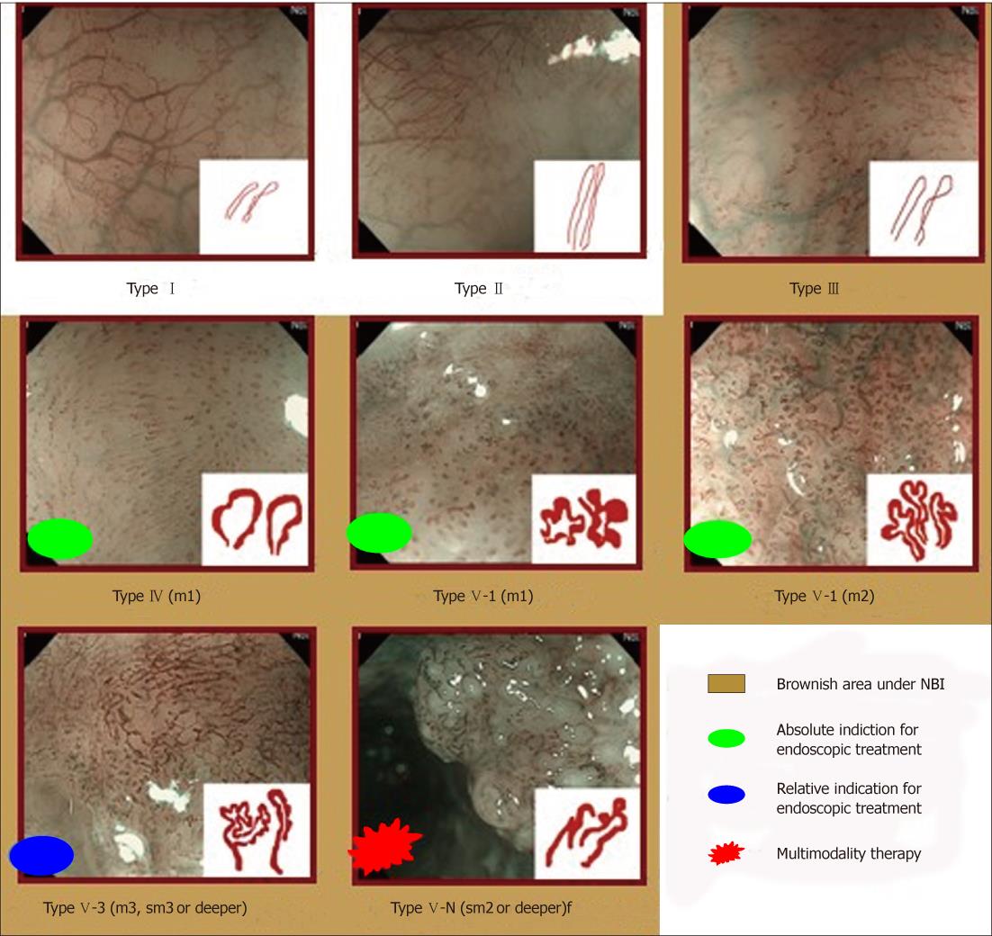
Figure 4 The case examples of Inoue’s intraepithelial papillary capillary loop classification from type I to type V-N.
NBI: Narrow-band imaging.
Figure 5 The morphology of Arima’s intraepithelial papillary capillary loop classification in esophagus.
LIN: Low-grade intraepithelial neoplasia; HIN: High-grade intraepithelial neoplasia.
Figure 6 The Paris classification of early lesion of gastrointestinal tract (A) and the depth of tumor infiltration (B).
ep: Epithelium; lmp: Lamina propria; mm: Muscularis mucosa; sm: Submucosa; mp:Muscularis propria.
- Citation: Chai NL, Ling-Hu EQ, Morita Y, Obata D, Toyonaga T, Azuma T, Wu BY. Magnifying endoscopy in upper gastroenterology for assessing lesions before completing endoscopic removal. World J Gastroenterol 2012; 18(12): 1295-1307
- URL: https://www.wjgnet.com/1007-9327/full/v18/i12/1295.htm
- DOI: https://dx.doi.org/10.3748/wjg.v18.i12.1295


