Copyright
©2011 Baishideng Publishing Group Co.
World J Gastroenterol. Jul 21, 2011; 17(27): 3184-3191
Published online Jul 21, 2011. doi: 10.3748/wjg.v17.i27.3184
Published online Jul 21, 2011. doi: 10.3748/wjg.v17.i27.3184
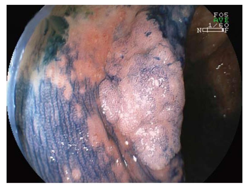
Figure 1 Visualization of a dysplasia-associated lesion or mass after topical application of indigo carmine.
Surface analysis revealed a Kudo pit pattern 3L.
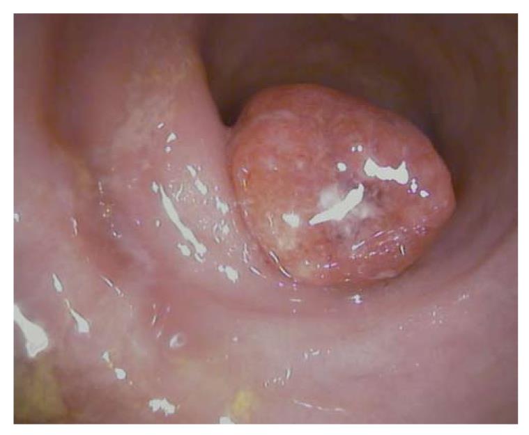
Figure 2 High-resolution standard white light endoscopic image of an adenoma-like mass in a patient with long standing ulcerative colitis.
Surface of the polyp is irregular and shows fibrin plaques. Histopathological analysis revealed high-grade intraepithelial neoplasia.
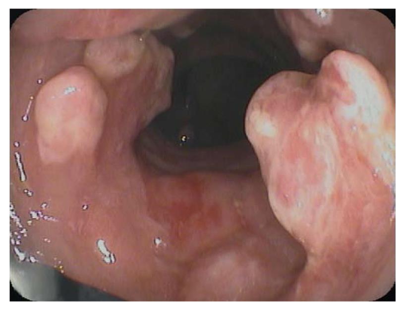
Figure 3 Multiple inflammatory polyps in chronic ulcerative colitis.
Image was recorded using the Pentax endomicroscope (EC-3870CIFK). Note the confocal lens at the 7 o’clock position.
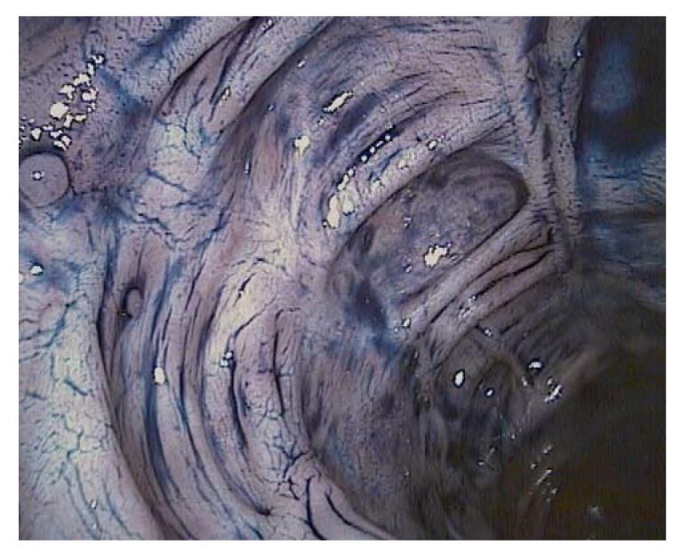
Figure 4 Chromoendoscopy with indigo carmine allows distinct surface analysis and demarcation of subtle lesions in long standing ulcerative colitis.
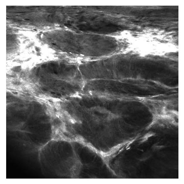
Figure 5 Fluorescein-guided confocal laser endomicroscopy (iCLE, Pentax, Tokyo, Japan) of dysplasia-associated lesion or mass.
Endomicroscopy visualizes tubular architecture and enlarged cells with depletion of goblet cells. The shape and size of the crypts is irregular, and leakage, demonstrated by the extravasation of fluorescein, is visible.
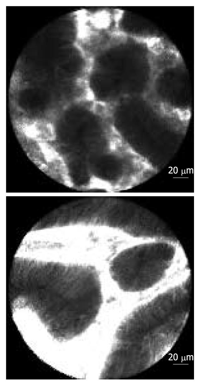
Figure 6 Fluorescein guided confocal laser endomicroscopy of adenoma-like mass (ALM; pCLE, Cellvizio, Mauna Kea Technologies, Paris, France).
Endomicroscopy shows villous transformation of colonic architecture and depletion of goblet cells indicating adenomatous tissue.
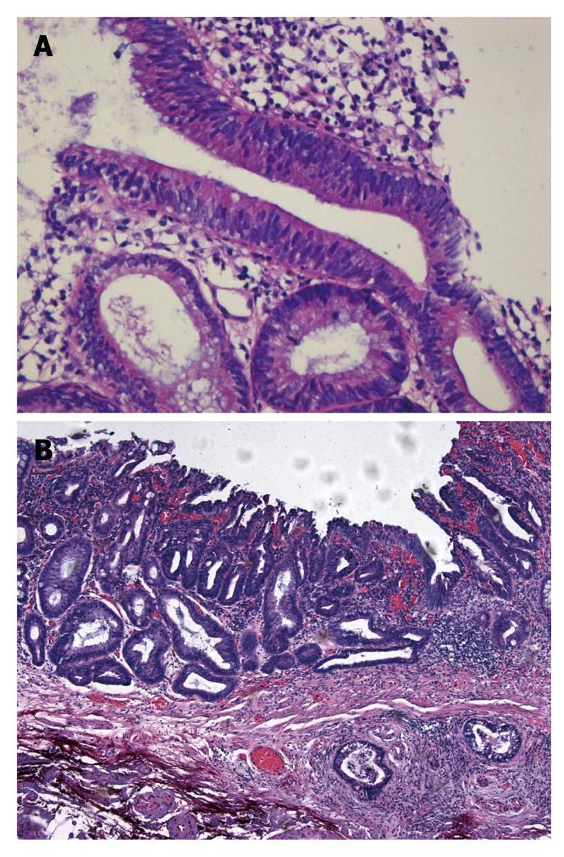
Figure 7 Histopathological image of dysplasia-associated lesion or mass with low-grade intraepithelial neoplasia in a patient with quiescent ulcerative colitis (A).
Panel B illustrates colitis-associated cancer with submucosal invasion.
- Citation: Neumann H, Vieth M, Langner C, Neurath MF, Mudter J. Cancer risk in IBD: How to diagnose and how to manage DALM and ALM. World J Gastroenterol 2011; 17(27): 3184-3191
- URL: https://www.wjgnet.com/1007-9327/full/v17/i27/3184.htm
- DOI: https://dx.doi.org/10.3748/wjg.v17.i27.3184


