
















©2011 Baishideng Publishing Group Co.
World J Gastroenterol. Jul 21, 2011; 17(27): 3184-3191
Published online Jul 21, 2011. doi: 10.3748/wjg.v17.i27.3184
Published online Jul 21, 2011. doi: 10.3748/wjg.v17.i27.3184
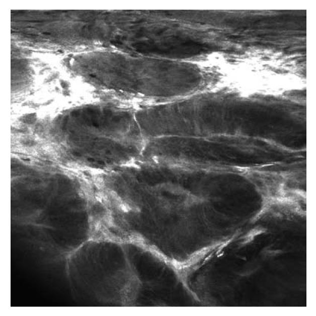
Figure 5 Fluorescein-guided confocal laser endomicroscopy (iCLE, Pentax, Tokyo, Japan) of dysplasia-associated lesion or mass.
Endomicroscopy visualizes tubular architecture and enlarged cells with depletion of goblet cells. The shape and size of the crypts is irregular, and leakage, demonstrated by the extravasation of fluorescein, is visible.
- Citation: Neumann H, Vieth M, Langner C, Neurath MF, Mudter J. Cancer risk in IBD: How to diagnose and how to manage DALM and ALM. World J Gastroenterol 2011; 17(27): 3184-3191
- URL: https://www.wjgnet.com/1007-9327/full/v17/i27/3184.htm
- DOI: https://dx.doi.org/10.3748/wjg.v17.i27.3184