Copyright
©2011 Baishideng Publishing Group Co.
World J Gastroenterol. Mar 21, 2011; 17(11): 1507-1514
Published online Mar 21, 2011. doi: 10.3748/wjg.v17.i11.1507
Published online Mar 21, 2011. doi: 10.3748/wjg.v17.i11.1507
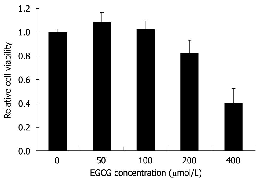
Figure 1 The cytotoxic effect of Epigallocatechin gallate on HepG2.
117 cells. Twenty four hours after seeding, cells were incubated with medium containing different concentrations of Epigallocatechin gallate (EGCG) for 3 d, and cell viability was determined by the 3-(4,5-Dimethylthiazol-2-yl)-2,5-diphenyltetrazolium bromide assay. Cell viability of the control well not treated with EGCG was determined and arbitrarily designated as 1. Error bars indicate standard deviation of 3 independent experiments.
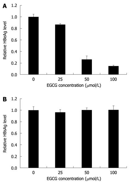
Figure 2 Epigallocatechin gallate inhibited hepatitis B virus e antigen expression level but not hepatitis B virus surface antigen expression level in the HepG2.
117 cell line. Twenty four hours after seeding, cells were incubated with medium containing different concentrations of Epigallocatechin gallate (EGCG) for 3 d. The hepatitis B virus e antigen (HBeAg) and hepatitis B virus surface antigen (HBsAg) expression levels in the supernatants were measured using enzyme-linked immunosorbent assay kits for measuring HBeAg and HBsAg, respectively. The level of HBeAg (A) and HBsAg (B) from cells not treated with EGCG (the control) were arbitrarily designated as 1. The levels of HBeAg (A) and HBsAg (B) in EGCG-treated cells were normalized with the control and expressed as relative levels. Error bars indicate standard deviation of 3 independent experiments.
Figure 3 Epigallocatechin gallate inhibited the production of hepatitis B virus precore mRNA.
Twenty four hours after seeding, HepG2.117 cells were treated with medium containing different concentration of Epigallocatechin gallate (EGCG) for 3 d. Total RNAs were prepared and were quantitated by semi-quantitative reverse transcription polymerase chain reaction (RT-PCR) with a pair of precore mRNA selective primers. Glyceraldehyde 3-phosphate dehydrogenase (GAPDH) mRNA was semi-quantitatively RT-PCR amplified and used for normalization. NC: Total RNA sample was subjected to PCR without reverse transcriptase and used as the negative control.
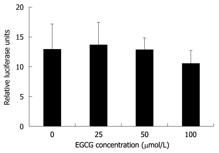
Figure 4 Epigallocatechin gallate does not affect hepatitis B virus core promoter activity.
Twelve hours after cotransfection with pHBVCP-Luc and pRL-TK, HepG2.117 cells were treated with media containing different concentrations of Epigallocatechin gallate (EGCG) for 3 d, and were lysed for the dual luciferase reporter analysis. Core promoter activity was normalized with control TK promoter activity and presented as relative luciferase units. Error bars indicate standard deviation of 3 independent experiments.
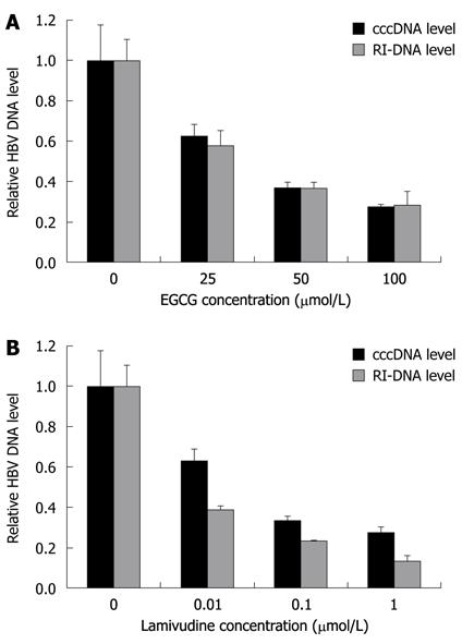
Figure 5 Epigallocatechin gallate down-regulates hepatitis B virus covalently closed circular DNA and replicative intermediates of DNA level.
A: Epigallocatechin gallate (EGCG) treatment was the same as described in Figure 1. Hepatitis B virus (HBV) covalently closed circular DNA (cccDNA) and total cellular HBV DNA were extracted and subjected to real-time polymerase chain reaction analysis; B: Lamivudine, the potent HBV DNA synthesis inhibitor, was used as a control inhibitor in this analysis. DNA level in the no treatment control was arbitrarily designated as 1. Error bars indicate standard deviation of 3 independent experiments. RI-DNA: Replicative intermediates of DNA.
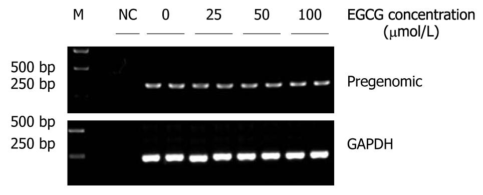
Figure 6 Epigallocatechin gallate did not inhibit hepatitis B virus pregenomic RNA transcription.
When doxycycline was removed from the cell culture medium, different concentrations of Epigallocatechin gallate (EGCG) were added to the HepG2.117 cells. After 3 d of culture, total RNA were extracted from the treated cells and the pregenomic RNAs (pgRNAs) were analyzed by semi-quantitative reverse transcription polymerase chain reaction. Glyceraldehyde 3-phosphate dehydrogenase (GAPDH) mRNAs were used for normalization. NC: Total RNA sample was subjected to polymerase chain reaction without reverse transcription.
- Citation: He W, Li LX, Liao QJ, Liu CL, Chen XL. Epigallocatechin gallate inhibits HBV DNA synthesis in a viral replication - inducible cell line. World J Gastroenterol 2011; 17(11): 1507-1514
- URL: https://www.wjgnet.com/1007-9327/full/v17/i11/1507.htm
- DOI: https://dx.doi.org/10.3748/wjg.v17.i11.1507


