Copyright
©2010 Baishideng.
World J Gastroenterol. Feb 28, 2010; 16(8): 1031-1033
Published online Feb 28, 2010. doi: 10.3748/wjg.v16.i8.1031
Published online Feb 28, 2010. doi: 10.3748/wjg.v16.i8.1031
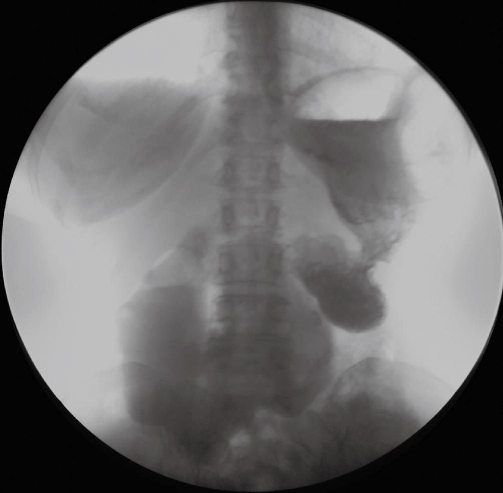
Figure 1 Two free communicating cavities can be seen just beyond the esophagogastric junction.
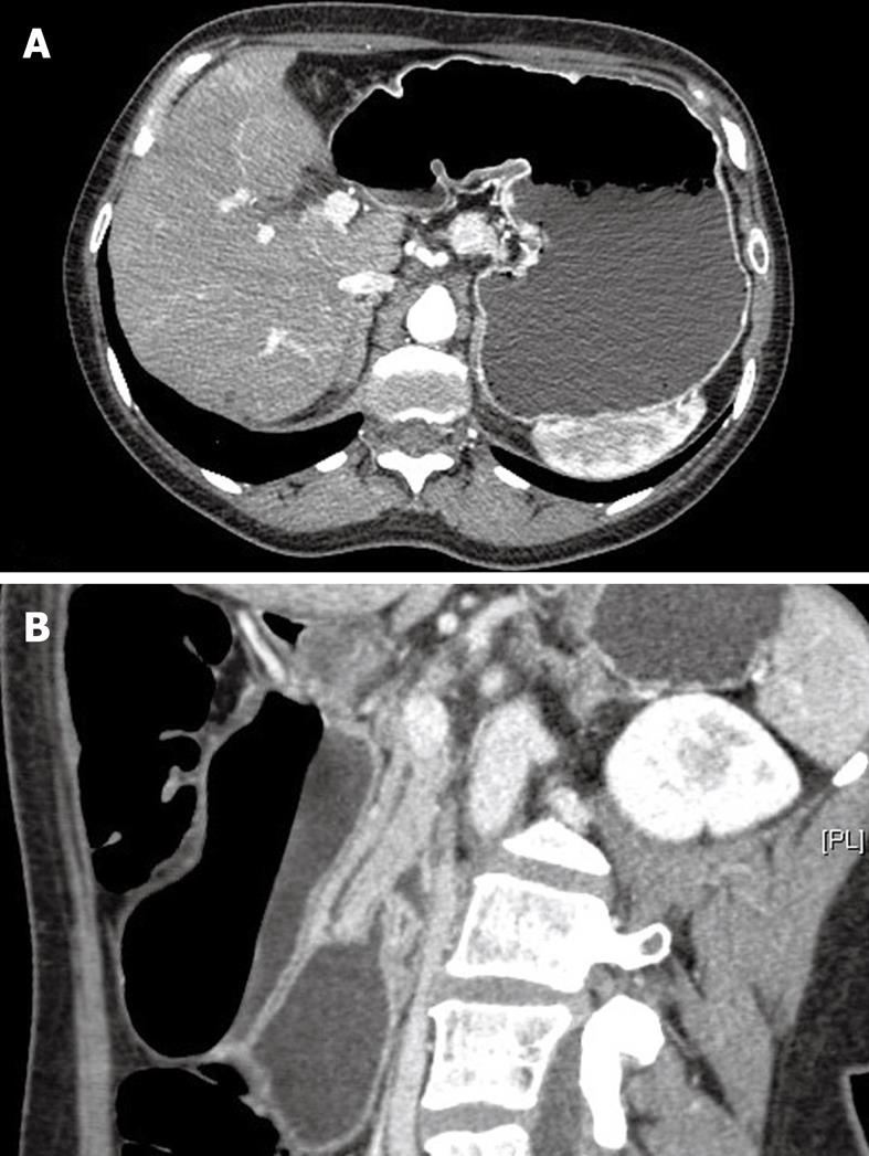
Figure 2 The communication of the 2 cavities visualized by computed tomography.
A: Frontal view; B: Lateral view.
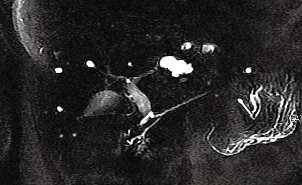
Figure 3 Magnetic resonance imaging showing the presence of an incomplete pancreas divisum.
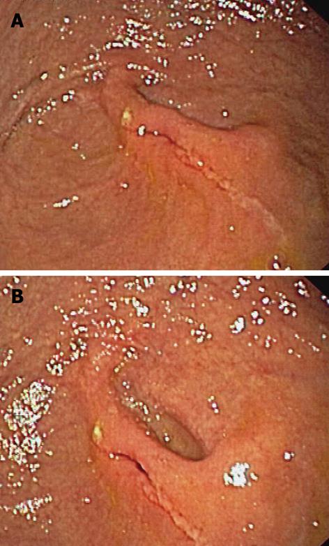
Figure 4 The endoscopic view of the duplication.
A: The first cavity with a similar gastric appearance continuing into the second one; B: The second proper gastric cavity continuing into the duodenum.
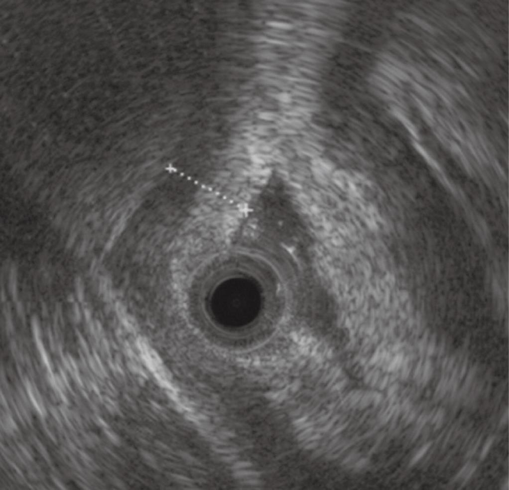
Figure 5 The typical gastric layer is visualized on endoscopic ultrasound.
- Citation: Di Pisa M, Curcio G, Marrone G, Milazzo M, Spada M, Traina M. Gastric duplication associated with pancreas divisum diagnosed by a multidisciplinary approach before surgery. World J Gastroenterol 2010; 16(8): 1031-1033
- URL: https://www.wjgnet.com/1007-9327/full/v16/i8/1031.htm
- DOI: https://dx.doi.org/10.3748/wjg.v16.i8.1031


