Copyright
©2010 Baishideng.
World J Gastroenterol. Feb 28, 2010; 16(8): 1025-1030
Published online Feb 28, 2010. doi: 10.3748/wjg.v16.i8.1025
Published online Feb 28, 2010. doi: 10.3748/wjg.v16.i8.1025
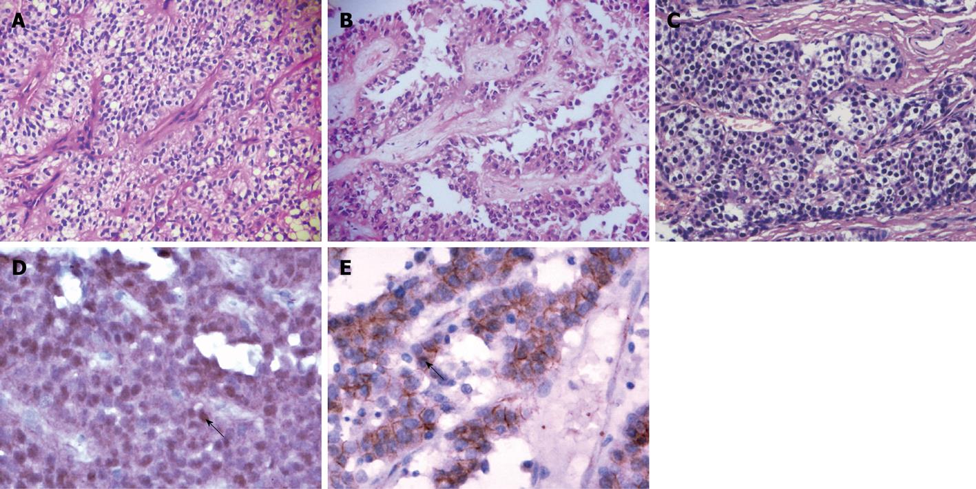
Figure 1 Histopathological and immunohistochemical features of solid-pseudopapillary neoplasm (SPN) and pancreatic endocrine tumor (PET).
A: SPN arranged in solid areas, patternless sheets of uniform epithelial cells with numerous small blood vessels (hematoxylin-eosin, original magnification × 200); B: SPN formed characteristic pseudopapillary changes due to the degenerative and discohesive nature of the tumor cells (hematoxylin-eosin, original magnification × 200); C: PET arranged in acinar-like pattern; the tumor cells are small and round with granular eosinophilic or clear cytoplasm (hematoxylin-eosin, original magnification × 200); D: β-catenin immunostaining of SPN: Nuclear translocation and accumulation of β-catenin protein (arrow) is seen in neoplastic epithelial cells (original magnification × 200); E: β-catenin immunostaining of PET: Membrane and cytoplasmic positive expression of β-catenin protein without nuclear stain (arrow) (original magnification × 200).
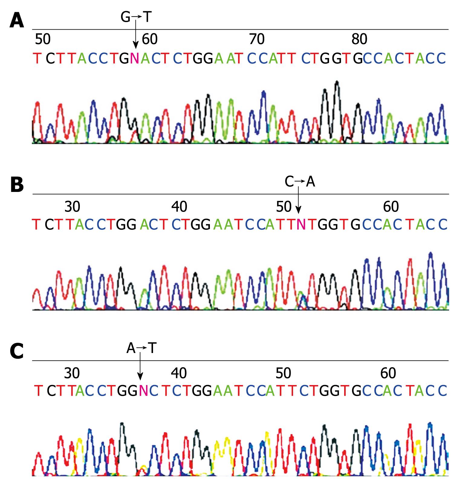
Figure 2 β-catenin oncogene mutations in SPN.
Representative DNA sequencing chromatograms demonstrate. A: A GAC→TAC mutation in codon 32 of case 5; B: A TCT→TAT mutation in codon 37 of case 2; C: A GAC→GTC mutation in codon 32 of case 9. Three samples of PET cases were randomly selected to be sequenced and the results confirmed that there was not any mutation on amplified PCR fragments of β-catenin gene exon 3.
- Citation: Liu BA, Li ZM, Su ZS, She XL. Pathological differential diagnosis of solid-pseudopapillary neoplasm and endocrine tumors of the pancreas. World J Gastroenterol 2010; 16(8): 1025-1030
- URL: https://www.wjgnet.com/1007-9327/full/v16/i8/1025.htm
- DOI: https://dx.doi.org/10.3748/wjg.v16.i8.1025


