Copyright
©2010 Baishideng.
World J Gastroenterol. Aug 7, 2010; 16(29): 3674-3679
Published online Aug 7, 2010. doi: 10.3748/wjg.v16.i29.3674
Published online Aug 7, 2010. doi: 10.3748/wjg.v16.i29.3674
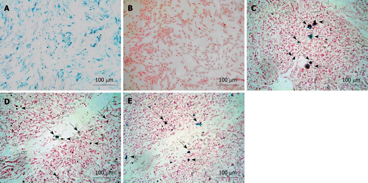
Figure 1 Characterization of labeled mesenchymal stem cells.
A: Almost 100% of labeled mesenchymal stem cells (MSCs) were positive for Prussian blue staining (magnification 100 ×); B: No blue particles were observed in unlabeled group (magnification 100 ×); C-E: Prussian blue staining for liver tissue slicing displayed several blue-positive cells scattering in and around sinusoids on day 3 and 7, and the experimental group at the endpoint of the experiment (magnification 100 ×). Arrows indicate Prussian blue positive MSCs.
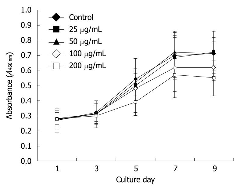
Figure 2 Growth curve of CCK-8 with Feridex-poly-l-lysine-labeled mesenchymal stem cells.
Cellular proliferation of 25 and 50 μg/mL subgroups were not significantly influenced (P > 0.05). Mesenchymal stem cells (MSCs) labeled with higher concentrations of complex were somewhat inhibited, which indicated that < 50 μg/mL Feridex-poly-l-lysine would be suitable for MSC labeling.
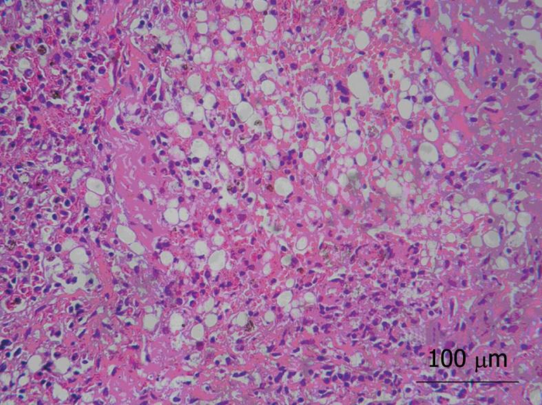
Figure 3 Histology of acutely injured liver tissue.
Liver tissue samples from the injured model group after 24 h injection of D-galactosamine demonstrated severe hepatic necrosis of 60%-70% of the lobule, sinusoidal congestion, vacuolization, trabecular fragmentation, and granulocytic infiltration of the portal space and septa (magnification 100 ×).
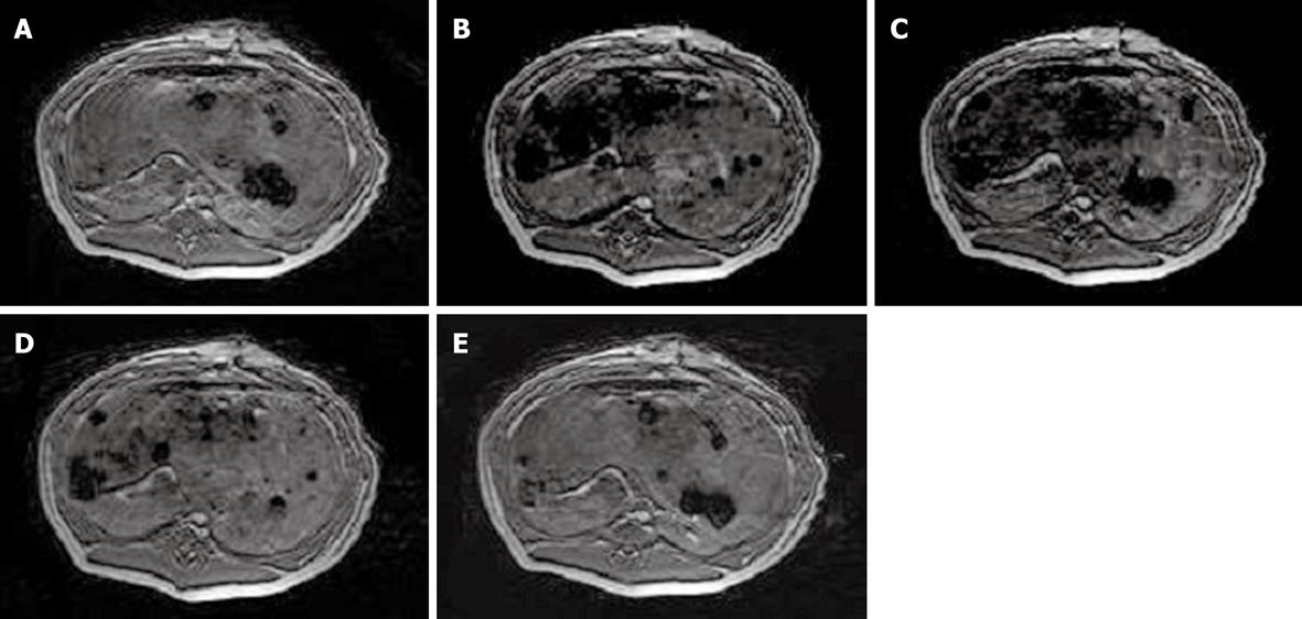
Figure 4 Magnetic resonance imaging of Feridex-poly-l-lysine labeled mesenchymal stem cells in the liver.
A: No loss of signal in the liver before transplantation; B: Significant signal intensity loss was observed in labeled mesenchymal stem cells on FFE sequences 6 h after transplantation; C, D: Attenuation of signal loss appeared over time; E: T2*-weighted images gradually approached to the normal level at the endpoint of the study.
- Citation: Shi XL, Gu JY, Han B, Xu HY, Fang L, Ding YT. Magnetically labeled mesenchymal stem cells after autologous transplantation into acutely injured liver. World J Gastroenterol 2010; 16(29): 3674-3679
- URL: https://www.wjgnet.com/1007-9327/full/v16/i29/3674.htm
- DOI: https://dx.doi.org/10.3748/wjg.v16.i29.3674


