Copyright
©2010 Baishideng.
World J Gastroenterol. Aug 7, 2010; 16(29): 3674-3679
Published online Aug 7, 2010. doi: 10.3748/wjg.v16.i29.3674
Published online Aug 7, 2010. doi: 10.3748/wjg.v16.i29.3674
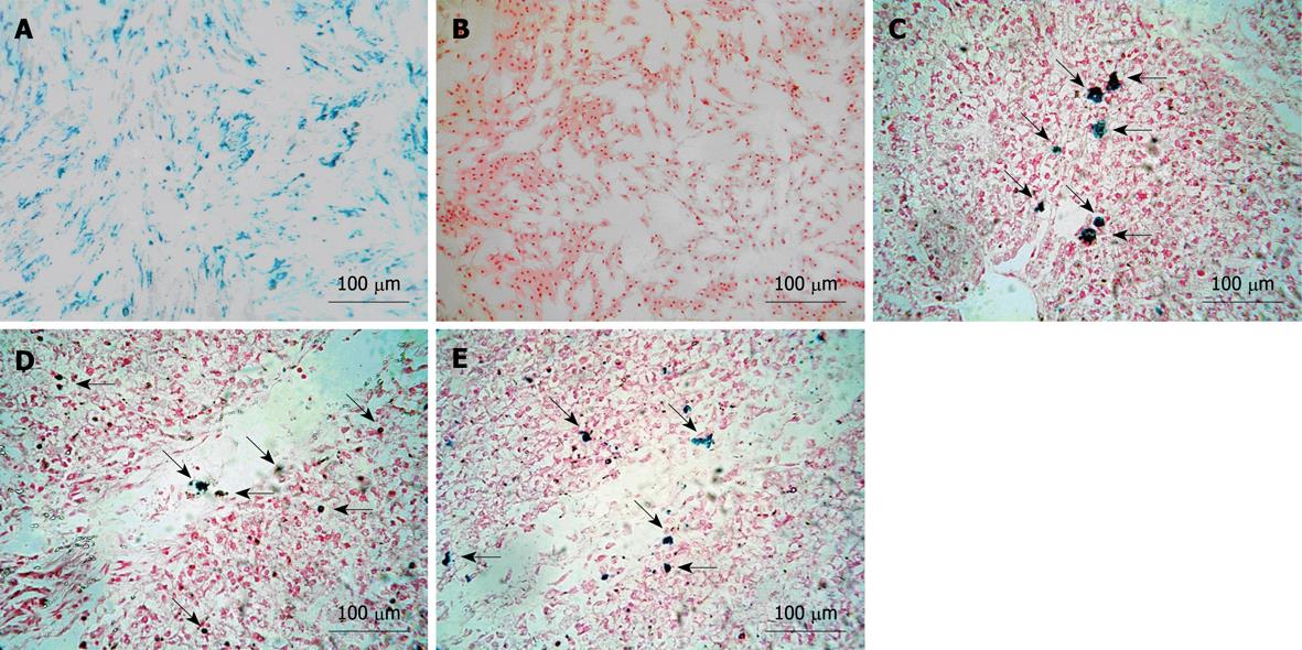
Figure 1 Characterization of labeled mesenchymal stem cells.
A: Almost 100% of labeled mesenchymal stem cells (MSCs) were positive for Prussian blue staining (magnification 100 ×); B: No blue particles were observed in unlabeled group (magnification 100 ×); C-E: Prussian blue staining for liver tissue slicing displayed several blue-positive cells scattering in and around sinusoids on day 3 and 7, and the experimental group at the endpoint of the experiment (magnification 100 ×). Arrows indicate Prussian blue positive MSCs.
- Citation: Shi XL, Gu JY, Han B, Xu HY, Fang L, Ding YT. Magnetically labeled mesenchymal stem cells after autologous transplantation into acutely injured liver. World J Gastroenterol 2010; 16(29): 3674-3679
- URL: https://www.wjgnet.com/1007-9327/full/v16/i29/3674.htm
- DOI: https://dx.doi.org/10.3748/wjg.v16.i29.3674


