Copyright
©2010 Baishideng.
World J Gastroenterol. Jun 7, 2010; 16(21): 2692-2697
Published online Jun 7, 2010. doi: 10.3748/wjg.v16.i21.2692
Published online Jun 7, 2010. doi: 10.3748/wjg.v16.i21.2692
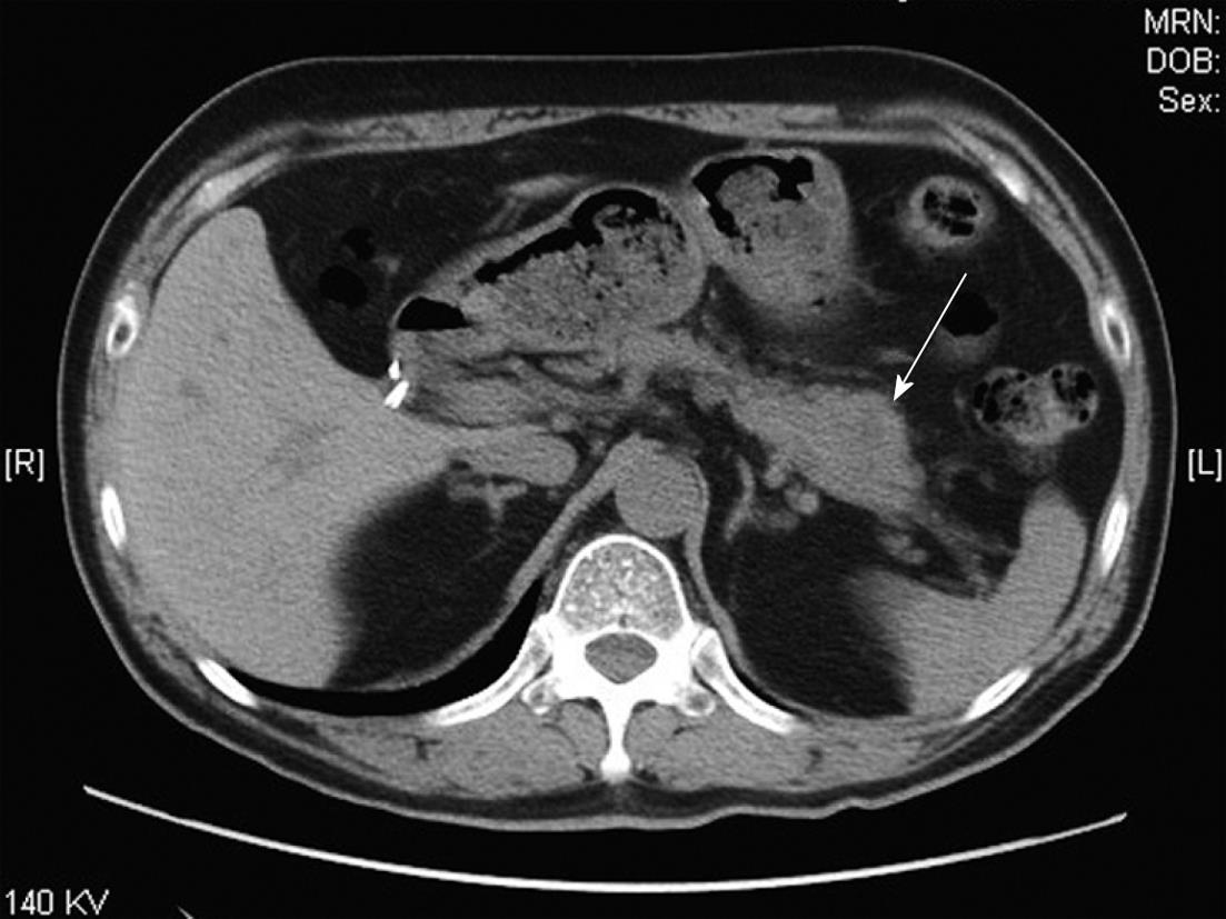
Figure 1 Computed tomography (CT) scan demonstrating a 3.
7 cm × 2.1 cm tumor (arrow) in pancreatic tail.
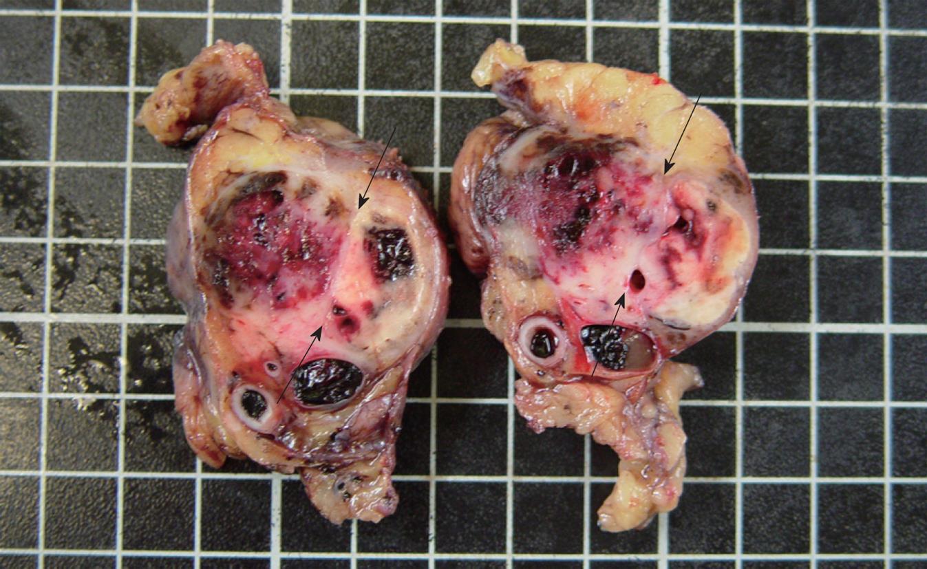
Figure 2 Gross examination showing a well-circumscribed tumor with a central fibrous band (arrows) separating the right exocrine and the left endocrine parts.
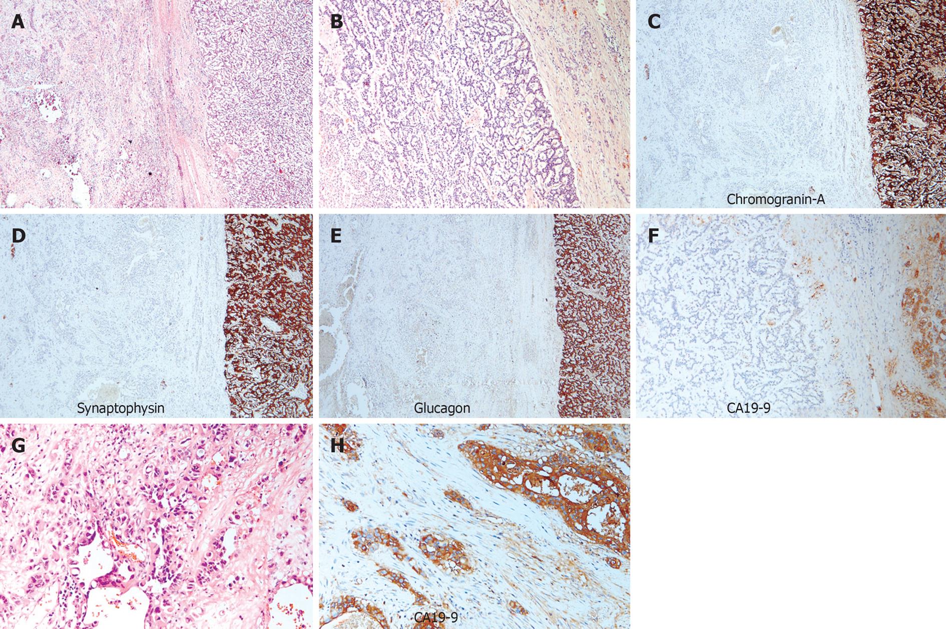
Figure 3 Microscopic features of solitary concomitant endocrine tumor and ductal adenocarcinoma of pancreas.
A: Low-power view showing ductal adenocarcinoma (left) and endocrine tumor (right) with a fibrous band (central) clearly demarcating the two components (HE × 40); B: Pancreatic endocrine tumor composed of monotonous small, round tumor cells arranged in gyriform or trabecullar patterns, marginated by a fibrous wall (right) (HE × 100); C-E: Pancreatic endocrine tumor (right) showing positive expression while ductal adenocarcinoma (left) showing negative expression of chromogranin A, synaptophysin and glucagon (IHC × 40); F: Pancreatic endocrine tumor (left) showing negative expression while residual benign pancreatic glands (right) showing positive expression of carbohydrate antigen 19-9 (CA19-9) (IHC × 100); G and H: Higher magnifications of ductal adenocarcinoma showing bizarre pleomorphic glands (HE × 200) and immunoreactive to CA19-9 (IHC × 200).
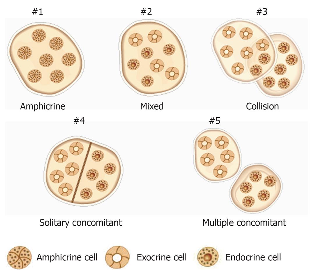
Figure 4 Five types of pancreatic neoplasm with combined exocrine and endocrine features.
- Citation: Chang SM, Yan ST, Wei CK, Lin CW, Tseng CE. Solitary concomitant endocrine tumor and ductal adenocarcinoma of pancreas. World J Gastroenterol 2010; 16(21): 2692-2697
- URL: https://www.wjgnet.com/1007-9327/full/v16/i21/2692.htm
- DOI: https://dx.doi.org/10.3748/wjg.v16.i21.2692


