Copyright
©2009 The WJG Press and Baishideng.
World J Gastroenterol. Oct 21, 2009; 15(39): 4976-4979
Published online Oct 21, 2009. doi: 10.3748/wjg.15.4976
Published online Oct 21, 2009. doi: 10.3748/wjg.15.4976
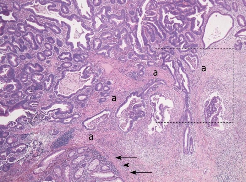
Figure 1 Low power view of an early invasive rectal adenocarcinoma with bundles of eosinophilic stromal cells (a), which are continuous with the muscularis mucosa (arrows, HE stain, original magnification × 40).
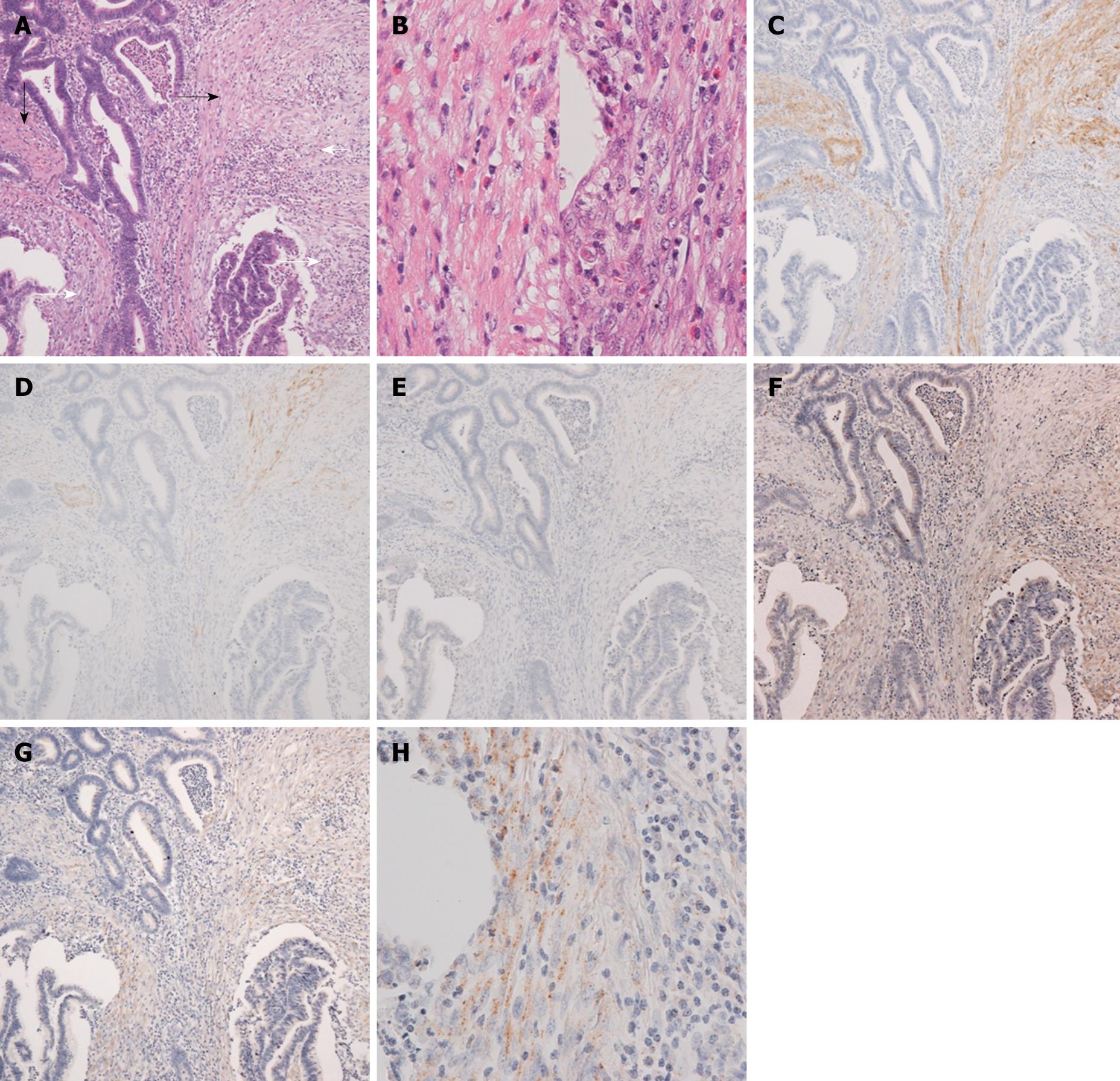
Figure 2 Middle power view of the invasive area (A) indicated by dotted box in Figure 1 shows a transition from bundles of eosinophilic stromal cells more similar to smooth muscle cells of the muscularis mucosa (black arrows) to those composed of less eosinophilic stromal cells with plumper nuclei (white arrows) (HE stain, × 100).
Higher magnification of the former eosinophilic stromal cells (left) and the latter ones (right) is shown in B (HE stain, × 400). Immunohistochemistry of the serial sections demonstrates the former being more constantly positive for α-SMA compared to the latter (C), and h-CD is positive in a few cells in the former, and negative in the latter (D). Desmin is almost negative for both the former and the latter (E). Immunohistochemical expression of type I procollagen (F) and the presence of type I procollagen mRNA by in situ hybridization (G, H) are demonstrated in the latter area (original magnification of C to G, × 100). Higher magnification of type I procollagen in situ hybridization is shown in H, which demonstrates positive signals in plump stromal cells (H, × 400).
- Citation: Ban S, Shimizu M. Muscularis mucosae in desmoplastic stroma formation of early invasive rectal adenocarcinoma. World J Gastroenterol 2009; 15(39): 4976-4979
- URL: https://www.wjgnet.com/1007-9327/full/v15/i39/4976.htm
- DOI: https://dx.doi.org/10.3748/wjg.15.4976


