Copyright
©2009 The WJG Press and Baishideng.
World J Gastroenterol. Sep 28, 2009; 15(36): 4593-4595
Published online Sep 28, 2009. doi: 10.3748/wjg.15.4593
Published online Sep 28, 2009. doi: 10.3748/wjg.15.4593
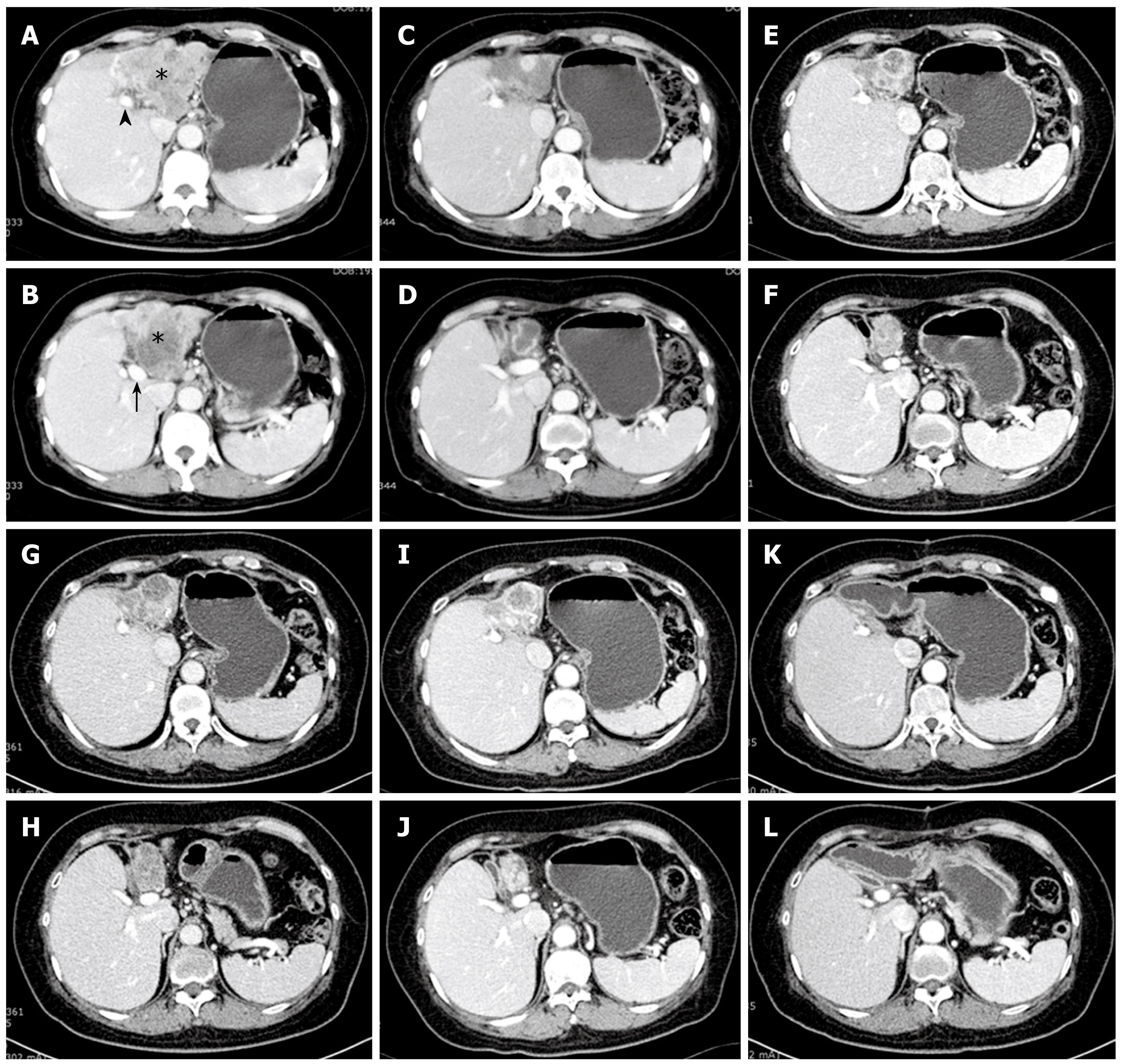
Figure 1 Abdominal CT scan images at two different levels are shown serially.
A: At left portal vein level (black arrowhead), a 10 cm sized irregular hypovascular mass (asterisk) occupying left hepatic lobe is shown; B: At lower, main portal vein level (black arrow), the inferiorly grown mass is shown. Invasion of middle hepatic vein and left portal vein were strongly suggested with multiple lymph node enlargements at the liver hilum and lesser omentum; C and D: On CT scan taken after the end of the first 6 cycles (18 times) of gemcitabine chemotherapy, the tumor mass decreased in size dramatically, smaller than half of the previous diameter; E and F: On CT scan on 3 years and 3 mo from diagnosis, mass does not show any significant change except for a little sclerotic change; G and H: On CT scan on 4 years from diagnosis, tumor mass shows re-increment of the size than previous study; I and J: On follow up CT scan after second 5 cycles of gemcitabine chemotherapy, partial response is shown again with slight decrease of the mass size; K and L: On CT scan taken 11 months after R0 resection, there is no any recurrence or new lesion growing.
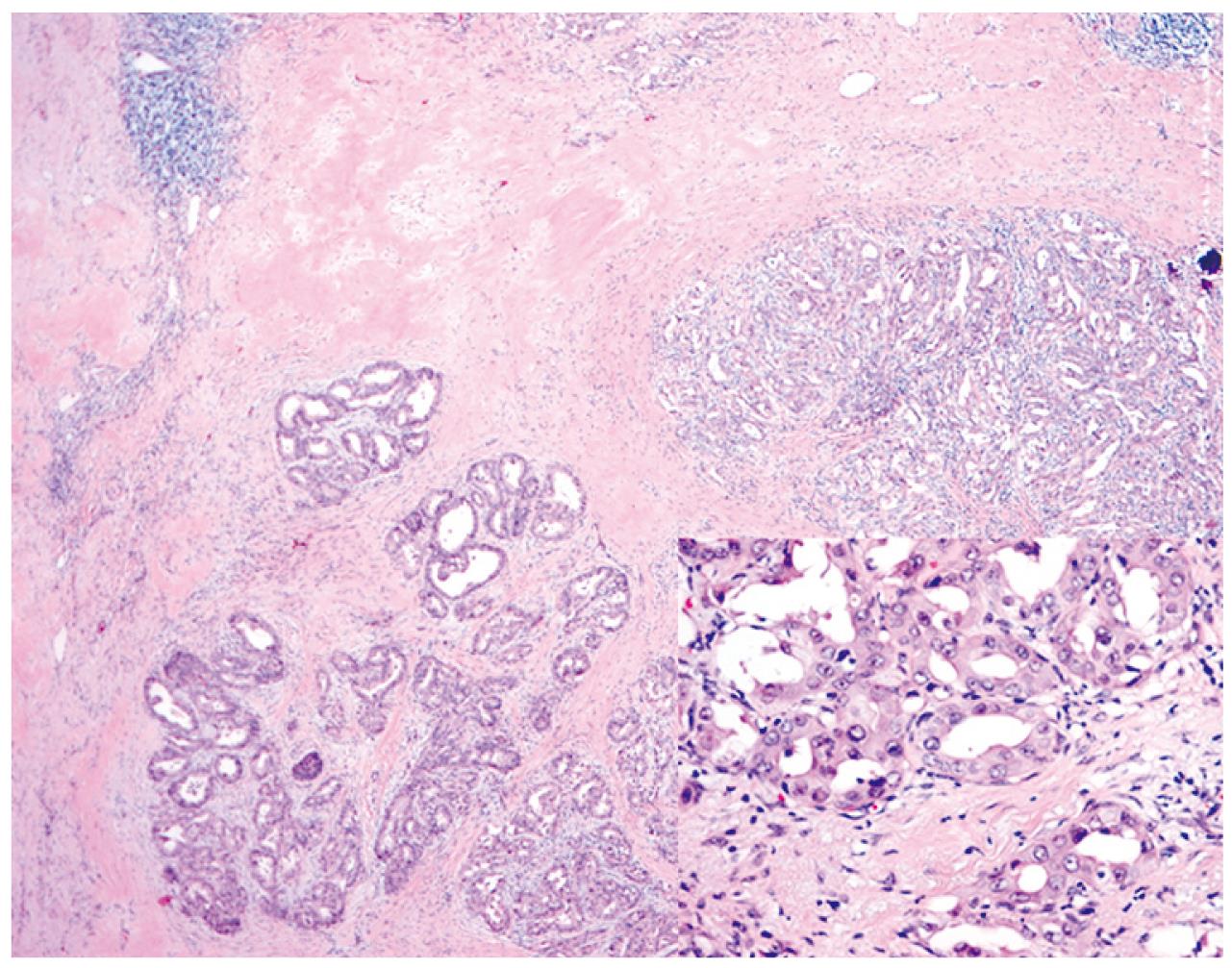
Figure 2 Histological finding of resected specimen is shown.
Well differentiated adenocarcinoma is seen with glandular structure and a dense fibrous stroma. Scattered lymphocytes are also present (40 × magnification). In higher magnification, tumor is composed of glands lined by cuboidal mucin-producing epithelium resemble biliary epithelium ( Inset, 400 × magnification).
- Citation: Kim SH, Kim IH, Kim SW, Lee SO. Repetitive response to gemcitabine that led to curative resection in cholangiocarcinoma. World J Gastroenterol 2009; 15(36): 4593-4595
- URL: https://www.wjgnet.com/1007-9327/full/v15/i36/4593.htm
- DOI: https://dx.doi.org/10.3748/wjg.15.4593


