Copyright
©2009 The WJG Press and Baishideng.
World J Gastroenterol. Sep 28, 2009; 15(36): 4593-4595
Published online Sep 28, 2009. doi: 10.3748/wjg.15.4593
Published online Sep 28, 2009. doi: 10.3748/wjg.15.4593
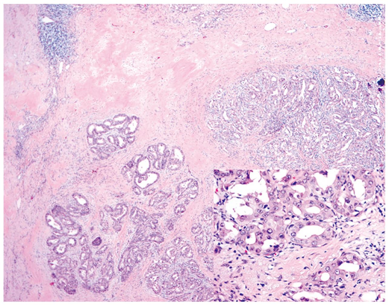
Figure 2 Histological finding of resected specimen is shown.
Well differentiated adenocarcinoma is seen with glandular structure and a dense fibrous stroma. Scattered lymphocytes are also present (40 × magnification). In higher magnification, tumor is composed of glands lined by cuboidal mucin-producing epithelium resemble biliary epithelium ( Inset, 400 × magnification).
- Citation: Kim SH, Kim IH, Kim SW, Lee SO. Repetitive response to gemcitabine that led to curative resection in cholangiocarcinoma. World J Gastroenterol 2009; 15(36): 4593-4595
- URL: https://www.wjgnet.com/1007-9327/full/v15/i36/4593.htm
- DOI: https://dx.doi.org/10.3748/wjg.15.4593


