Copyright
©2009 The WJG Press and Baishideng.
World J Gastroenterol. Jul 7, 2009; 15(25): 3191-3193
Published online Jul 7, 2009. doi: 10.3748/wjg.15.3191
Published online Jul 7, 2009. doi: 10.3748/wjg.15.3191
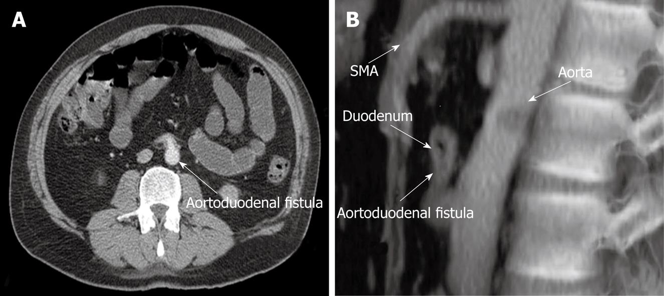
Figure 1 CT and lateral aortography.
A: CT showing direct extravasation of contrast material from the aorta into the duodenal lumen (arrow); B: Lateral aortography showing aortoduodenal fistula. Note there is no evidence of abdominal aneurysm.
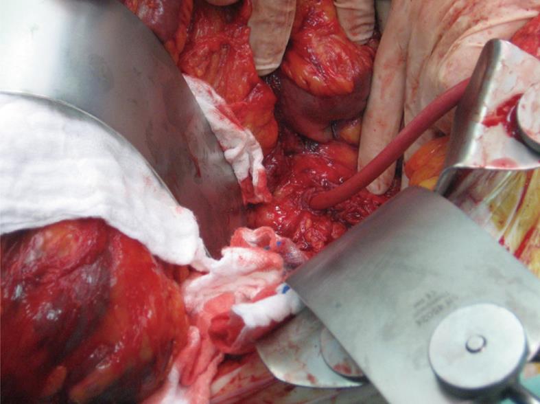
Figure 2 Perioperative findings of primary fistula between the third part of the duodenum and aorta.
A Foley catheter was inserted to control the bleeding.
- Citation: Bala M, Sosna J, Appelbaum L, Israeli E, Rivkind AI. Enigma of primary aortoduodenal fistula. World J Gastroenterol 2009; 15(25): 3191-3193
- URL: https://www.wjgnet.com/1007-9327/full/v15/i25/3191.htm
- DOI: https://dx.doi.org/10.3748/wjg.15.3191


