Copyright
©2009 The WJG Press and Baishideng.
World J Gastroenterol. Apr 14, 2009; 15(14): 1779-1781
Published online Apr 14, 2009. doi: 10.3748/wjg.15.1779
Published online Apr 14, 2009. doi: 10.3748/wjg.15.1779
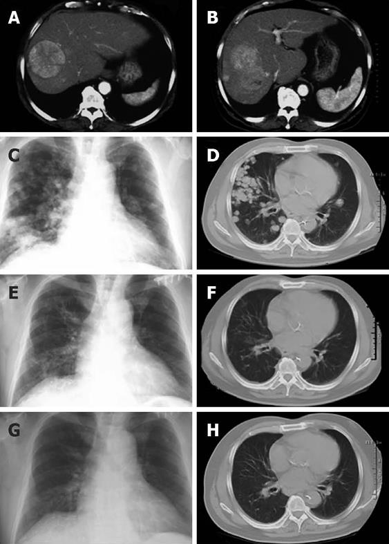
Figure 1 Chest X-ray and CT images before and after chemotherapy (A-H).
Seven months before the chemotherapy, huge multiple HCCs were apparent in the right lobe of the liver (A, B). On admission before chemotherapy, multiple lung metastases were seen in the bilateral lung fields (C, D). Two months after starting chemotherapy, the multiple lung metastases had disappeared completely (E, F). Five months after starting chemotherapy, the tumors had not recurred (G, H).
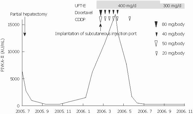
Figure 2 Clinical course of this patient.
First, 400 mg/d of oral UFT-E was started. We then placed an intra-arterial catheter that delivered medication into the aorta just before the bronchial arteries, and docetaxel (80 mg/body initially, followed by 40 mg/body) and CDDP (50 mg/body initially, followed by 20 mg/body) were administered every 2 wk via a subcutaneous injection port. Levels of PIVKA-II, a tumor marker, decreased rapidly 1 mo after starting chemotherapy; levels continued to fall to normal and were maintained.
- Citation: Tsuchiya A, Imai M, Kamimura H, Togashi T, Watanabe K, Seki KI, Ishikawa T, Ohta H, Yoshida T, Kamimura T. Successful treatment of multiple lung metastases of hepatocellular carcinoma by combined chemotherapy with docetaxel, cisplatin and tegafur/uracil. World J Gastroenterol 2009; 15(14): 1779-1781
- URL: https://www.wjgnet.com/1007-9327/full/v15/i14/1779.htm
- DOI: https://dx.doi.org/10.3748/wjg.15.1779


