Copyright
©2009 The WJG Press and Baishideng.
World J Gastroenterol. Mar 14, 2009; 15(10): 1259-1263
Published online Mar 14, 2009. doi: 10.3748/wjg.15.1259
Published online Mar 14, 2009. doi: 10.3748/wjg.15.1259
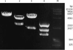
Figure 1 Restriction map of recombinant plasmid PCI-neo-aPKCα.
Lane 1: PCI-neo digested with SalI EcoR1; Lane 2: PCI-neo-aPKCα digested with BamH1; Lane 3: PCI-neo-aPKCα digested with Sa l; Lane 4: PCI-neo-aPKCα digested with BamH1 + SalI; Lane M: λ DNA HindIII Marker.
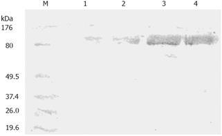
Figure 2 Western blot identification of PKCα protein in SGC7901, SGC7901/VCR, SGC7901/VCR/neo and SGC7901/VCR/aPKC cells.
Lane M: BenchmarkTM Marker; Lane 1: SGC7901 cells; Lane 2: SGC7901/VCR/aPKC cells; Lane 3: SGC7901/VCR/neo cells; Lane 4: SGC7901/VCR cells.
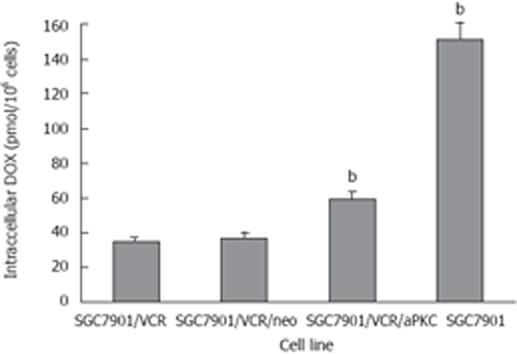
Figure 3 Accumulation of intracellular DOX in SGC7901, SGC7901/VCR, SGC7901/VCR/neo and SGC7901/VCR/aPKC cells.
Cells were incubated with 4.0 &mgr;mol/L DOX for 60 min. Each point represents the mean ± SD from three experiments. bP < 0. 01 vs SGC7901/VCR cells.
- Citation: Wu DL, Sui FY, Du C, Zhang CW, Hui B, Xu SL, Lu HZ, Song GJ. Antisense expression of PKCα improved sensitivity of SGC7901/VCR cells to doxorubicin. World J Gastroenterol 2009; 15(10): 1259-1263
- URL: https://www.wjgnet.com/1007-9327/full/v15/i10/1259.htm
- DOI: https://dx.doi.org/10.3748/wjg.15.1259


