Copyright
©2009 The WJG Press and Baishideng.
World J Gastroenterol. Mar 14, 2009; 15(10): 1246-1253
Published online Mar 14, 2009. doi: 10.3748/wjg.15.1246
Published online Mar 14, 2009. doi: 10.3748/wjg.15.1246
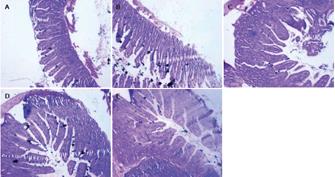
Figure 1 Comparison of morphological changes of small intestinal mucosa (including villous height, villous width, villous area, crypt depth, and ratio of villous height to crypt depth) at different development periods in SD rats (HE, x 100).
A: Group of 3 wk; B: Group of 5 wk; C: Group of 7 wk; D: Group of 9 wk; E: group of 11 wk.
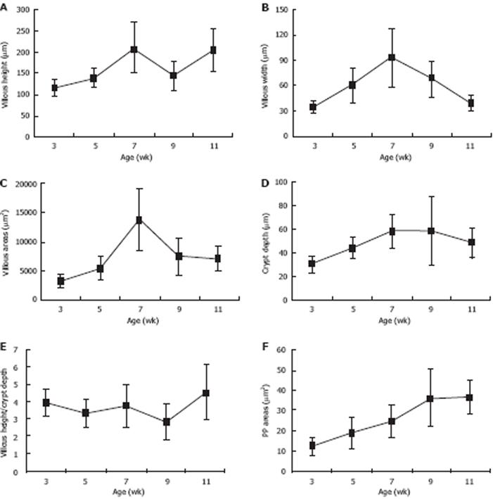
Figure 2 Morphological analysis of villous-crypt axis: All morphological parameters matured as age increased.
The results were presented as mean ± SD from 5 rats. A: Villous height increased at 3 wk postpartum, decreased 7 to 9 wk postpartum, and increased again after 9 wk postpartum; B: Villous width increased at 3 wk postpartum, peaked at 7 wk postpartum; C: Villous area increased significantly between 5 and 7 wk postpartum, peaked at 7 wk postpartum; D: Crypt depth increased from 3 to 7 wk postpartum and decreased slightly at 9 wk postpartum; E: Ratio of villous height to crypt depth were relatively stable and increased significantly from 9 to 11 wk postpartum; F: PP increased from 3 to 11 wk postpartum.
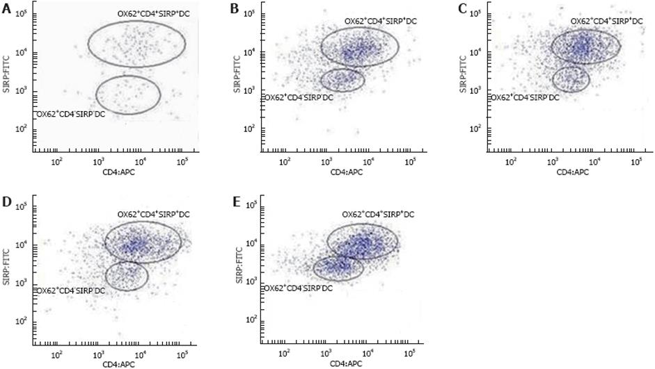
Figure 3 FCM of expression of OX62+CD4+SIRP+DC and OX62+CD4-SIRP-DC subsets at different development periods in SD rats.
A: Group of 3 wk; B: Group of 5 wk; C: Group of 7 wk; D: Group of 9 wk; E: Group of 11 wk.
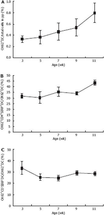
Figure 4 Flow cytometric analyses of OX62+CD4+SIRP+ and OX62+CD4-SIRP- PP DCs in vivo at 3, 5, 7, 9 and 11 wk postpartum.
The results were presented as mean ± SD from 5 rats. A: Significant growth occurred in different age groups for the number of OX62+DCs; B: Levels of OX62+CD4+OX41+DC subsets increased significantly at 3-5 wk postpartum and 7-9 wk postpartum; C: OX62+CD4-SIRP-DC levels declined on the whole, with small fluctuation from 7 to 9 wk postpartum.
- Citation: Zhou YJ, Gao J, Yang HM, Zhu JX, Chen TX, He ZJ. Morphology and ontogeny of dendritic cells in rats at different development periods. World J Gastroenterol 2009; 15(10): 1246-1253
- URL: https://www.wjgnet.com/1007-9327/full/v15/i10/1246.htm
- DOI: https://dx.doi.org/10.3748/wjg.15.1246


