Copyright
©2008 The WJG Press and Baishideng.
World J Gastroenterol. Feb 14, 2008; 14(6): 966-968
Published online Feb 14, 2008. doi: 10.3748/wjg.14.966
Published online Feb 14, 2008. doi: 10.3748/wjg.14.966
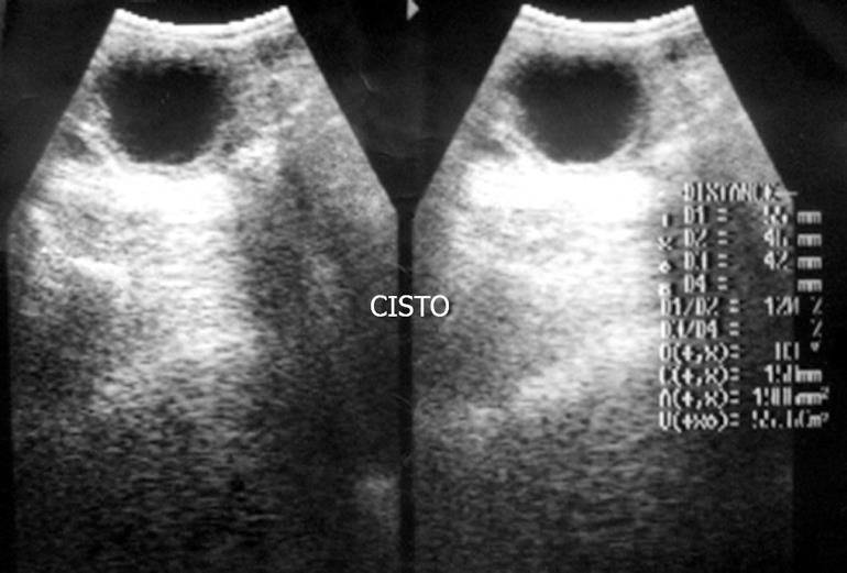
Figure 1 Ultrasonography demonstrating a cystic lesion.
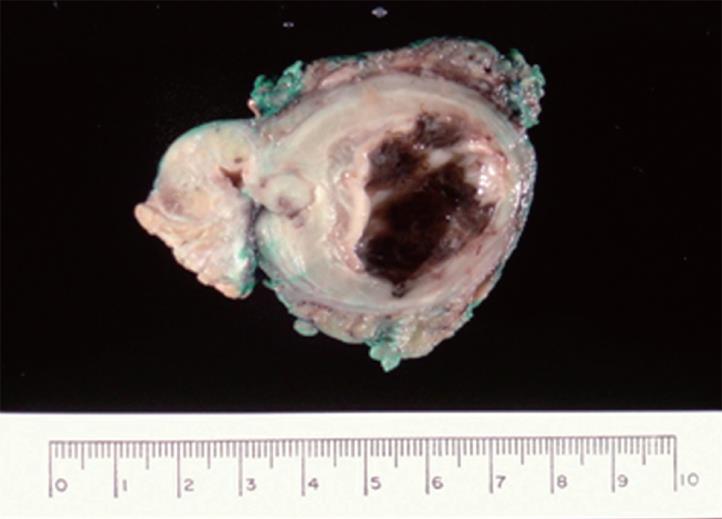
Figure 2 Surgical specimen including distal pancreas and the cyst.
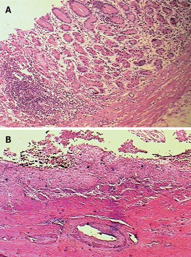
Figure 3 Histological section of the cyst wall.
A: Ectopic gastric mucosa with gastritis (HE, × 100); B: Ectopic gastric mucosa with ulceration (HE, × 25).
- Citation: Carneiro FP, Sobreira MN, Azevedo AEB, Alves APR, Campos KM. Colonic duplication in an adult mimicking a tumor of pancreas. World J Gastroenterol 2008; 14(6): 966-968
- URL: https://www.wjgnet.com/1007-9327/full/v14/i6/966.htm
- DOI: https://dx.doi.org/10.3748/wjg.14.966


