Copyright
©2008 The WJG Press and Baishideng.
World J Gastroenterol. Nov 21, 2008; 14(43): 6738-6742
Published online Nov 21, 2008. doi: 10.3748/wjg.14.6738
Published online Nov 21, 2008. doi: 10.3748/wjg.14.6738
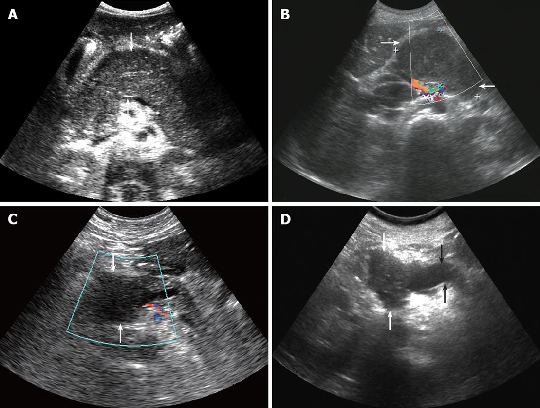
Figure 1 ultrasonic manifestation.
A: Diffuse pancreatic lymphoma (arrows): the pancreas showed diffuse enlargement and the echo decreased; B: Pancreatic lymphoma (arrows): a low echo lump was seen in the pancreatic head. There were no blood flow signals; C: Pancreatic cancer (arrows): a low echo lump was seen in the pancreatic head. There were no blood flow signals; D: Pancreatic cancer: a low echo lump appeared in the pancreatic head (white arrows) and the main pancreatic duct (black arrows) was obviously expanded.
- Citation: Qiu L, Luo Y, Peng YL. Value of ultrasound examination in differential diagnosis of pancreatic lymphoma and pancreatic cancer. World J Gastroenterol 2008; 14(43): 6738-6742
- URL: https://www.wjgnet.com/1007-9327/full/v14/i43/6738.htm
- DOI: https://dx.doi.org/10.3748/wjg.14.6738


