Copyright
©2008 The WJG Press and Baishideng.
World J Gastroenterol. Aug 7, 2008; 14(29): 4677-4683
Published online Aug 7, 2008. doi: 10.3748/wjg.14.4677
Published online Aug 7, 2008. doi: 10.3748/wjg.14.4677
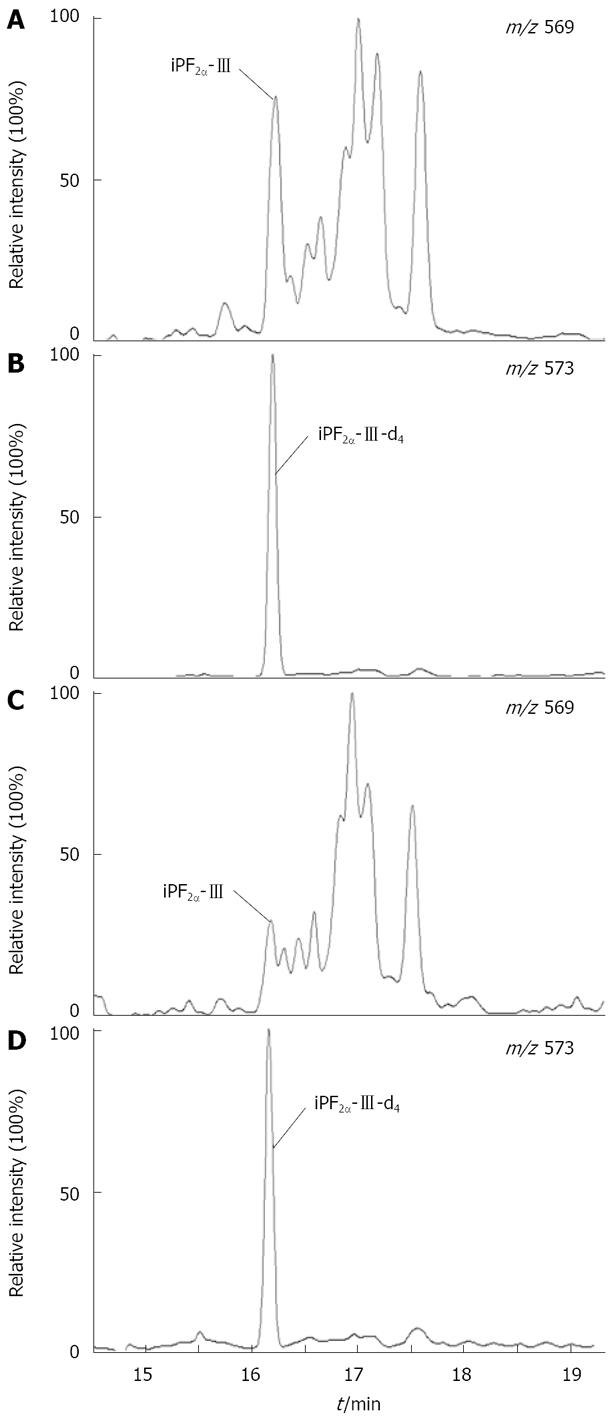
Figure 1 GC-NICI-MS SIM chromatograms of (A) plasma total iPF2α-III and (C) hepatic iPF2α-III isolated from a rat with simple steatosis, and (B) and (D) the internal standard iPF2α-III-d4 in the plasma and tissue samples, respectively.
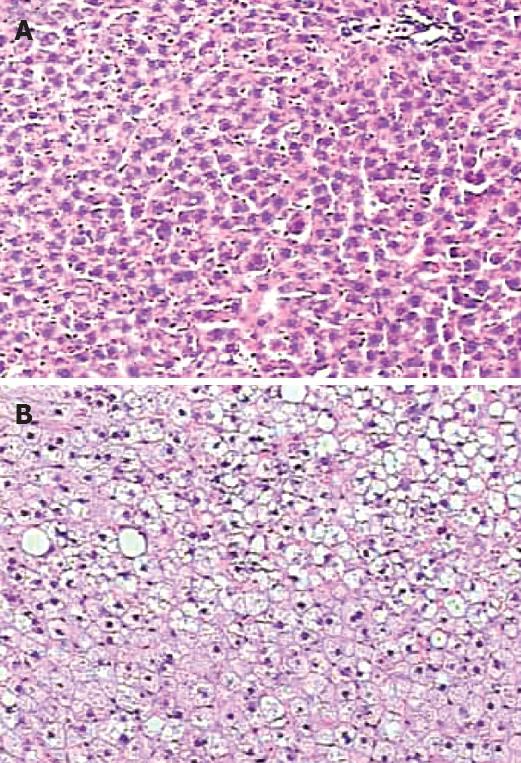
Figure 2 Enhanced lipid accumulation in the liver of rat by high-fat diet.
(A) Control, and (B) Steatotic liver (HE, magnification × 100).
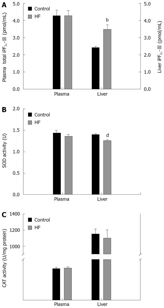
Figure 3 Plasma and hepatic levels of iPF2α-III and antioxidant enzymes in rats.
(A) Significant differences in iPF2α-III levels were found in the liver, but not in plasma between HF rats and control; (B) Significant decreases in SOD activity were found in the liver, but not in erythrocytes of HF rats compared to that in control, and (C) Reducing tendency of CAT activities was observed in liver, but not in blood of HF rats. bP < 0.01 and dP < 0.001.
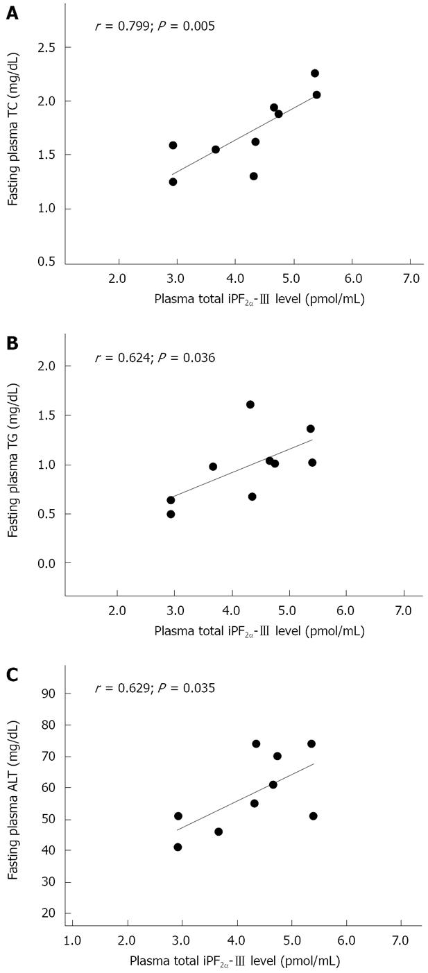
Figure 4 The formation of plasma total iPF2α-III was significantly correlated with the levels of fasting plasma TC (A), TG (B) and ALT (C) in HF rats.
- Citation: Zhu MJ, Sun LJ, Liu YQ, Feng YL, Tong HT, Hu YH, Zhao Z. Blood F2-isoprostanes are significantly associated with abnormalities of lipid status in rats with steatosis. World J Gastroenterol 2008; 14(29): 4677-4683
- URL: https://www.wjgnet.com/1007-9327/full/v14/i29/4677.htm
- DOI: https://dx.doi.org/10.3748/wjg.14.4677


