Copyright
©2008 The WJG Press and Baishideng.
World J Gastroenterol. Jun 21, 2008; 14(23): 3642-3649
Published online Jun 21, 2008. doi: 10.3748/wjg.14.3642
Published online Jun 21, 2008. doi: 10.3748/wjg.14.3642
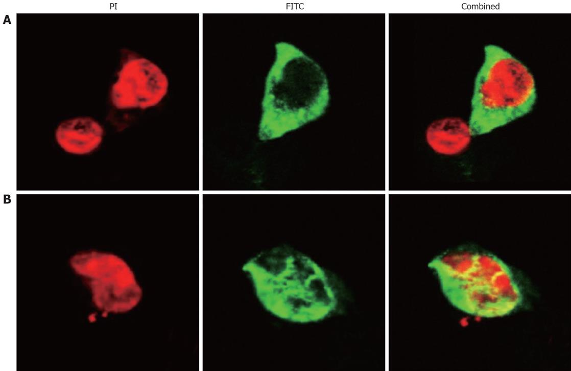
Figure 1 SP-TAT-apoptin expression in HepG2 cells (× 1000).
Cells transfected with plenti6/V5-D-TOPO/SP-TAT-apoptin plasmid and fixed at 24 h (A) and 48 h (B) post-transfection. Recombinant apoptin detected by anti-V5-FITC antibody is shown in green and cell nuclei stained by PI in red. Apoptin protein showed a diffuse pattern in the cytoplasm at 24 h post-transfection, and in the nucleus at 48 h.
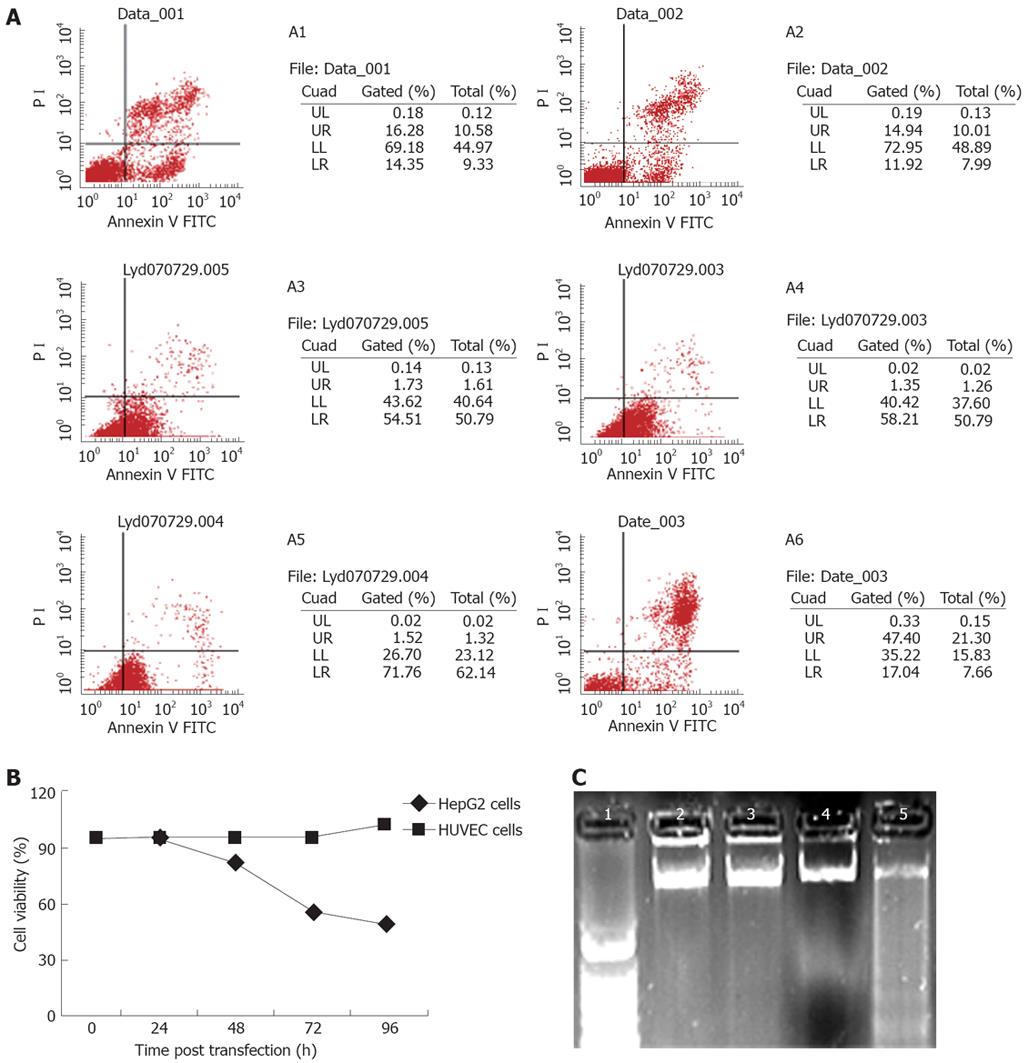
Figure 2 SP-TAT-apoptin-induced cell death.
A: Cell viability measured by flow cytometry. A1 and A2: HUVECs at 48 and 72 h post-transfection; A3-A6: HepG2 cells at 24, 48, 72 and 96 h. HepG2 cells were susceptible to SP-TAT-apoptin-induced apoptosis in a time-dependent manner; B: Cell viability determined by MTT dye reduction assay; C: DNA fragmentation in HepG2 cells demonstrated by agarose gel electrophoresis. Lane 1: 1 kb DNA marker; Lanes 2-5: DNA from cells at 24, 48, 60 and 72 h post-transfection, respectively.
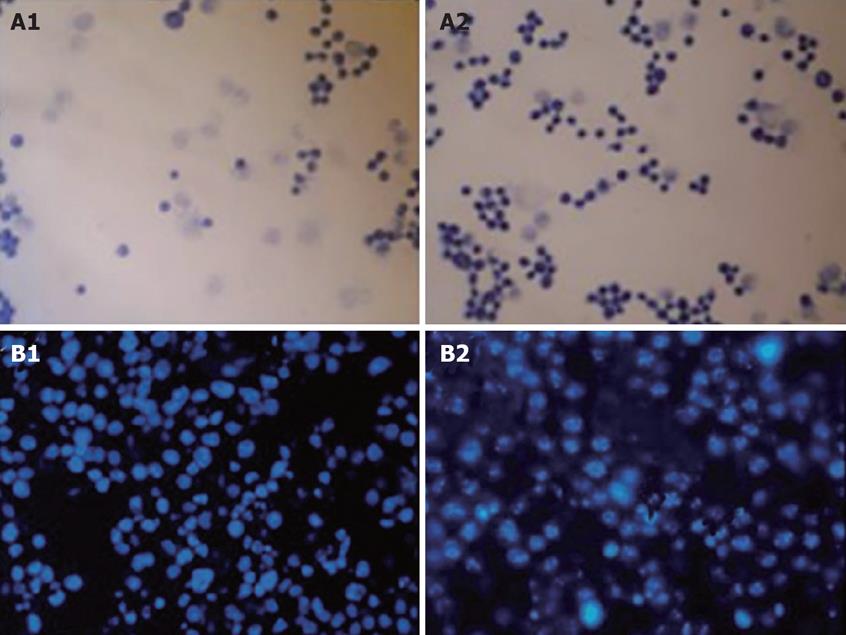
Figure 3 Cytotoxicity of SP-TAT-apoptin compared to SP-TAT.
A: Micrographs of HepG2 cells transfected with SP-TAT-apoptin construct stained by Apoptotic/Necrotic Cell Detection Kit. An inverted microscope (× 400) was used. The nuclei of apoptotic cells were stained deep blue. A1 and A2: HepG2 cells at 24 and 72 h post-transfection; B: HepG2 cells stained with DAPI and observed by fluorescence microscopy (× 400). B1: HepG2 cells 72 h after transfection with plenti6/V5-D-TOPO/SP-TAT-GFP plasmid; B2: HepG2 cells 72 h after transfection with plenti6/V5-D-TOPO/SP-TAT-apoptin. Arrow indicates apoptotic cells.
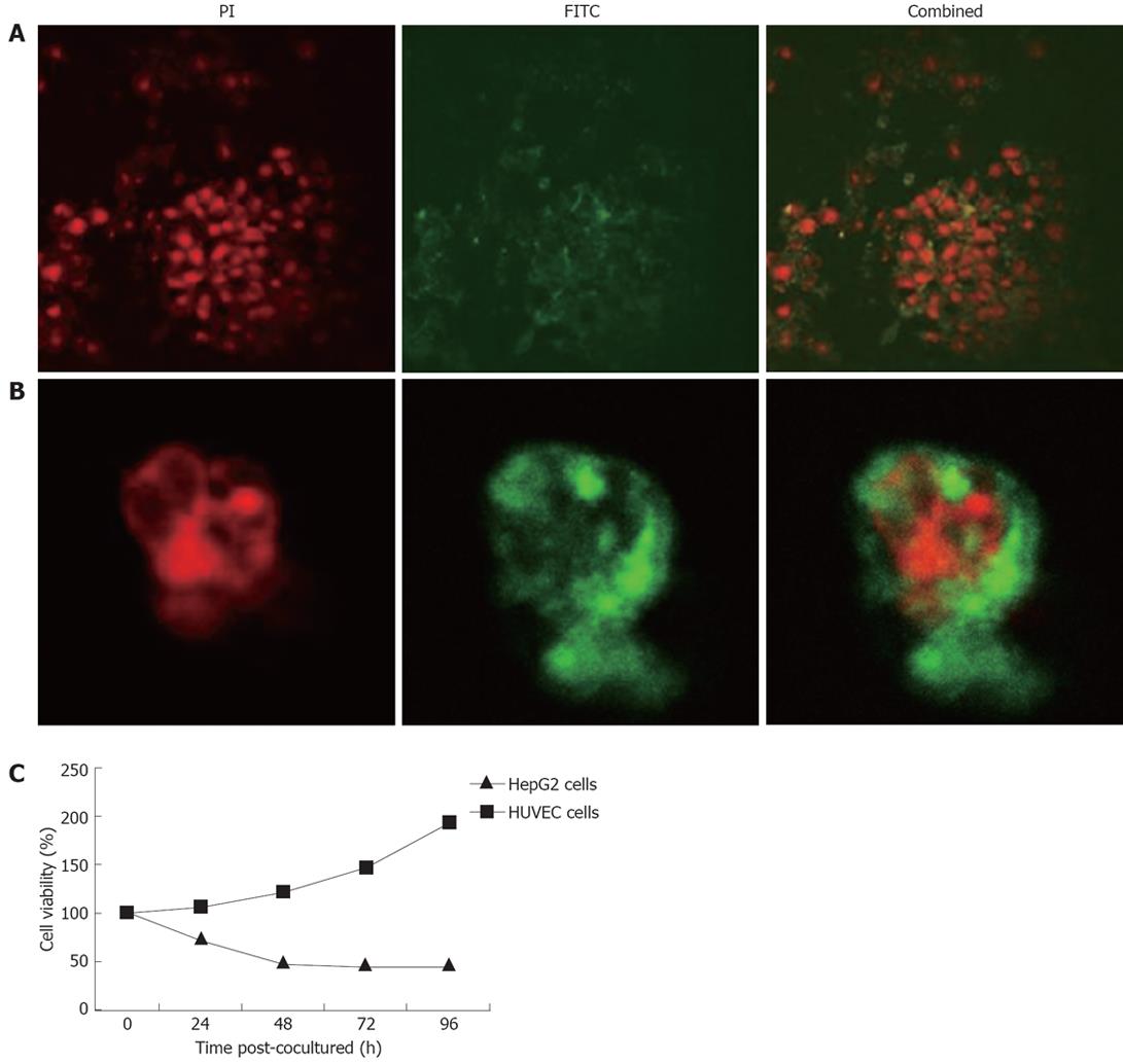
Figure 4 Cell death induced by secreted TAT-apoptin.
A: HepG2 cells immunostained 4 h after co-culture with the supernatant of CHO cells expressing SP-TAT-apoptin. Recombinant TAT-apoptin detected by anti-V5-FITC antibody is shown in green and cell nuclei stained by PI in red (× 1000); B: Translocation of recombinant apoptin in HepG2 cells 24 h after co-culture with the supernatant of CHO cell expressing SP-TAT-apoptin; C: Cell viability determined by MTT dye reduction assay after co-culture with secreted TAT-apoptin.
- Citation: Han SX, Ma JL, Lv Y, Huang C, Liang HH, Duan KM. Secretory Transactivating Transcription-apoptin fusion protein induces apoptosis in hepatocellular carcinoma HepG2 cells. World J Gastroenterol 2008; 14(23): 3642-3649
- URL: https://www.wjgnet.com/1007-9327/full/v14/i23/3642.htm
- DOI: https://dx.doi.org/10.3748/wjg.14.3642


