Copyright
©2008 The WJG Press and Baishideng.
World J Gastroenterol. Jun 7, 2008; 14(21): 3403-3409
Published online Jun 7, 2008. doi: 10.3748/wjg.14.3403
Published online Jun 7, 2008. doi: 10.3748/wjg.14.3403
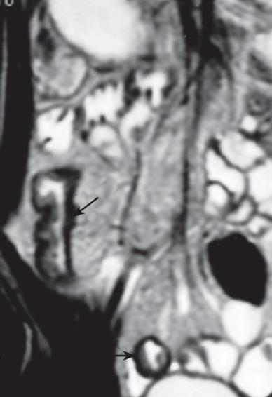
Figure 1 A 36-year-old man with Crohn's disease, the small bowel thickness exceeds 4-5 mm on T2W image, and stratified appearance (so-called "target" or "double halo" appearance) can be seen.
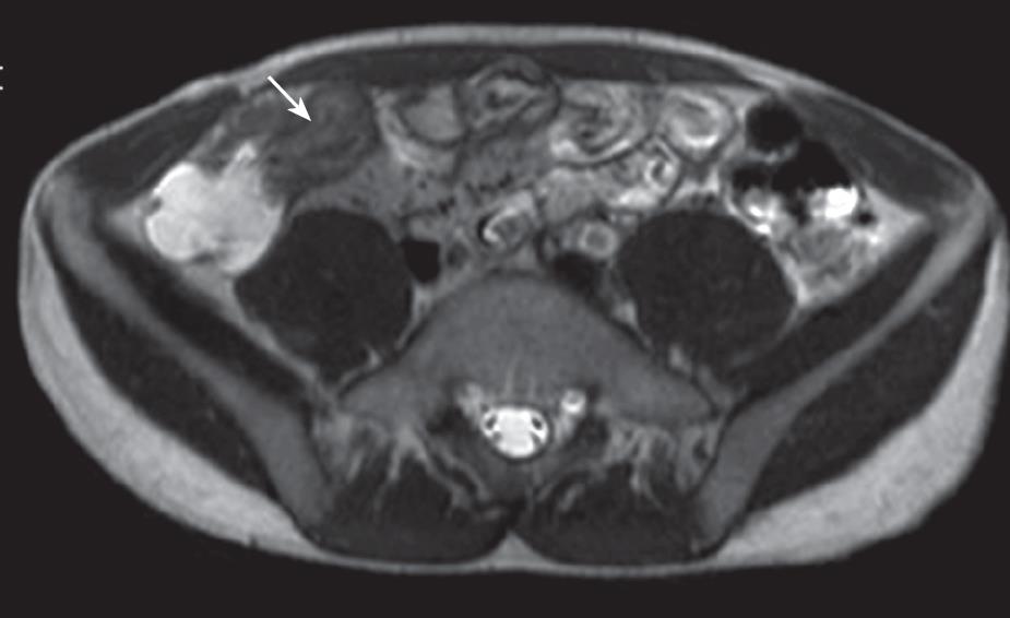
Figure 2 A 25-year-old man with Crohn's disease and inflammation of ileocecal junction.
T2W image shows "double halo" appearance (arrows) of thickened (8 mm) bowel wall.
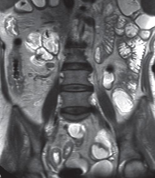
Figure 3 A 42-year-old man with Crohn's disease, T2W image shows skip lesions of ileum with thickened (7 mm) bowel wall.
Figure 4 A 36-year-old woman with Crohn's disease, the bowel wall of the involved segment has a homogeneous enhancement at CE-T1W image.
And the “comb sign” also can be seen.
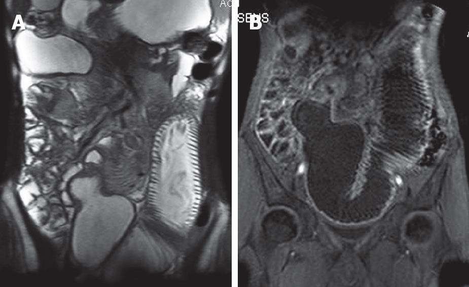
Figure 5 A: A 68-year-old man with adenocarcinomas, T2W image shows the tumor with similar signal intensity, the proximate jejunum dilating conspicuously; B: The same patient, tumor shows heterogeneous enhancement greater than adjacent bowel on gadolinium-enhanced image.
- Citation: Zhu J, Xu JR, Gong HX, Zhou Y. Updating magnetic resonance imaging of small bowel: Imaging protocols and clinical indications. World J Gastroenterol 2008; 14(21): 3403-3409
- URL: https://www.wjgnet.com/1007-9327/full/v14/i21/3403.htm
- DOI: https://dx.doi.org/10.3748/wjg.14.3403


