Copyright
©2008 The WJG Press and Baishideng.
World J Gastroenterol. Mar 28, 2008; 14(12): 1936-1940
Published online Mar 28, 2008. doi: 10.3748/wjg.14.1936
Published online Mar 28, 2008. doi: 10.3748/wjg.14.1936
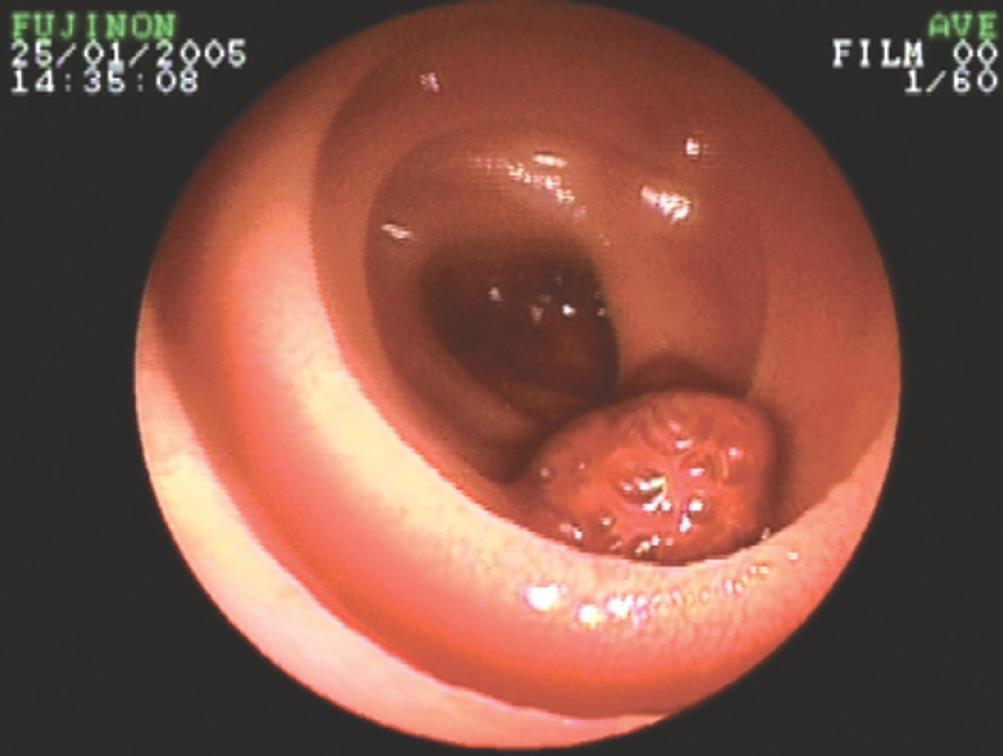
Figure 1 Enteroscopy identified a polyp 1.
5 cm in diameter in jejunum.
Figure 2 Stromal tumor of small intestine.
A: Preoperative enteroscopy; B: Surgical exploration; C: Photomicrograph of the lesion (HE, × 100).
Figure 3 Adenocarcinoma of small intestine.
A: Preoperative enteroscopy; B: Surgical exploration; C: Photomicrograph of the lesion (HE, × 100).
Figure 4 Diverticula was visualized as angeioma by preoperative double-balloon enteroscopy.
A: Preoperative enteroscopy, angeioma between jejunum and ileum; B: Diverticula 80 cm proximal to ileocecum. C: Photomicrograph of the lesion (HE, × 100).
Figure 5 Vascular malformation of small intestine.
A: Preoperative enteroscopy; B: Surgical exploration; C: Photomicrograph of the lesion (HE, × 100).
- Citation: Lin MB, Yin L, Li JW, Hu WG, Qian QJ. Double-balloon enteroscopy reliably directs surgical intervention for patients with small intestinal bleeding. World J Gastroenterol 2008; 14(12): 1936-1940
- URL: https://www.wjgnet.com/1007-9327/full/v14/i12/1936.htm
- DOI: https://dx.doi.org/10.3748/wjg.14.1936


