Copyright
©2008 The WJG Press and Baishideng.
World J Gastroenterol. Mar 14, 2008; 14(10): 1575-1581
Published online Mar 14, 2008. doi: 10.3748/wjg.14.1575
Published online Mar 14, 2008. doi: 10.3748/wjg.14.1575
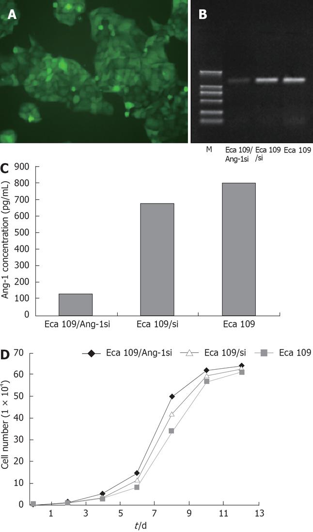
Figure 1 Effect of Ang-1si on Ang-1 expression in Eca109 esophageal cancer cells.
A: Eca109 cells were infected with Ad/Ang-1si at a multiplicity of infection (MOI) of 50, nearly 100% of cultured Eca109/Ang-1si cells were GFP-positive under the fluorescent microscope; B: Ang-1 mRNA was quantified by RT-PCR. Compared with Eca109, the Ang-1 mRNA level of Eca109/Ang-1si cells was reduced by 80%, but no significant alteration in Eca109/si cells; C: Secreted Ang-1 protein was quantified by ELISA. Compared with Eca109, the Ang-1 concentration in the media was decreased by Ad/Ang-1si, but no significant change in the cell line transfected with Ad/si; D: Cell number was measured at various time points, there was no difference of cell growth curve among Eca109, Eca109/si, and Eca109/Ang-1si cells.
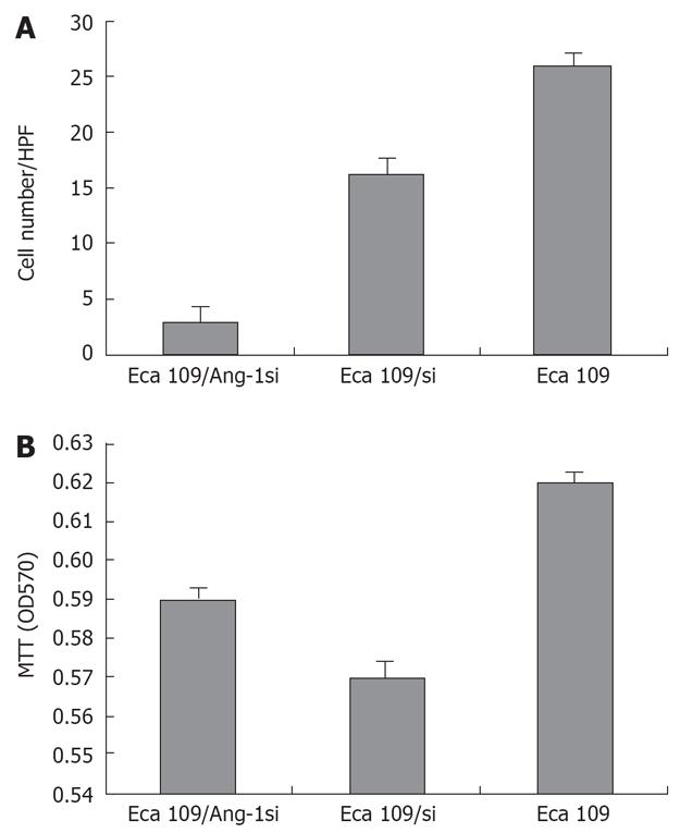
Figure 2 Effect of Ang-1si on HUVEC cell proliferation and chemotaxis.
A: HUVEC cell migration was quantified after 6 h, few HUVEC cells migrated as the chemotactic stimulus from Eca109/Ang-1si cells supernatants compared with Eca109 and Eca109/si cells supernatants. bP < 0.01; B: HUVEC cells were incubated with the indicated supernatant for 48 h, no difference of proliferative activity of Eca109/Ang-1si cells from Eca109 and Eca109/si cells. bP < 0.01.
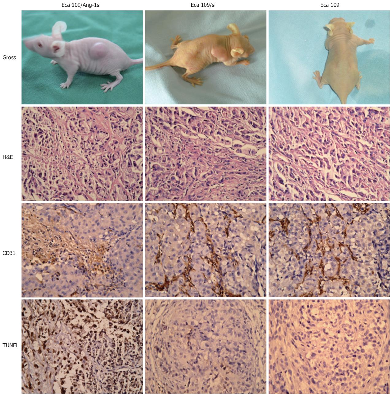
Figure 3 Effect of Ang-1si on esophageal cancer growth in vivo.
Grossly, the Eca109/Ang-1si tumors were significantly smaller than those in both control groups; Under microscopy, massive necrotic tissue was found in Eca109/Ang-1si tumors, while Eca109 and Eca109/si tumors showed fine necrosis; the Eca109/Ang-1si tumors were avascular with lower MVD, while Eca109 and Eca109/si tumors appeared very vascularized with higher MVD; Eca109/Ang-1si tumors underwent massive apoptosis with more TUNEL-positive cells compared with Eca109 and Eca109/si tumors, which showed a finely granular cytoplasm with evenly dispersed chromatin and lower TUNEL-positive cells.
- Citation: Liu XH, Bai CG, Yuan Y, Gong DJ, Huang SD. Angiopoietin-1 targeted RNA interference suppresses angiogenesis and tumor growth of esophageal cancer. World J Gastroenterol 2008; 14(10): 1575-1581
- URL: https://www.wjgnet.com/1007-9327/full/v14/i10/1575.htm
- DOI: https://dx.doi.org/10.3748/wjg.14.1575


